. 2005 Apr;88(4):2789-800.
doi: 10.1529/biophysj.104.046235. Epub 2005 Jan 14.

Assembly of lipoprotein particles containing apolipoprotein-B: structural model for the nascent lipoprotein particle

Affiliations

Assembly of lipoprotein particles containing apolipoprotein-B: structural model for the nascent lipoprotein particle

Paul E Richardson et al. Biophys J. 2005 Apr.

Abstract

Apolipoprotein B (apoB) is the major protein component of large lipoprotein particles that transport lipids and cholesterol. We have developed a detailed model of the first 1000 residues of apoB using standard sequence alignment programs (ClustalW and MACAW) and the MODELLER6 package for three-dimensional homology modeling. The validity of the apoB model was supported by conservation of disulfide bonds, location of all proline residues in turns and loops, and conservation of the hydrophobic faces of the two C-terminal amphipathic beta-sheets, betaA (residues 600-763) and betaB (residues 780-1000). This model suggests a lipid-pocket mechanism for initiation of lipoprotein particle assembly. In a previous model we suggested that microsomal triglyceride transfer protein might play a structural role in completion of the lipid pocket. We no longer think this likely, but instead propose a hairpin-bridge mechanism for lipid pocket completion. Salt-bridges between four tandem charged residues (717-720) in the turn of the hairpin-bridge and four tandem complementary residues (997-1000) at the C-terminus of the model lock the bridge in the closed position, enabling the deposition of an asymmetric bilayer within the lipid pocket.


Figures

FIGURE 1
FIGURE 1
Alignment and analysis of apoB-22.5. (A) Sequence alignment for the first 620 amino acids of human apoB and the homologous residues in lipovitellin. (B) The red boxes represent local sequence homology between apoB and lipovitellin that were recognized using the alignment program MACAW. (C) The yellow boxes are conserved regions in mouse and human apoB sequence that the program LOCATE identified as potential amphipathic β-strands.
FIGURE 2
FIGURE 2
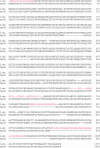
Alignment over the first 1000 residues of apoB. The complete alignment was used to make the apoB1000 model. The first 620 residues of the alignment were taken from Fig. 1 A. The alignment from 621–1000 was done using the results from the MACAW and LOCATE analyses (Fig. 1, B and C). The text highlighted in red represents residues not resolved in the lipovitellin crystal structure.
FIGURE 3
FIGURE 3
ApoB-1000. (A) RIBBONS diagram of the apoB1000 model looking from the large opening of the β-sheets toward the small opening. (B) RIBBONS diagram of the apoB1000 model looking from the small opening of the β-sheets (180° rotation from Fig. 4 A). Residues 670–745 are not shown, but the asterisk symbol (*) in A (left diagram) indicates where the loop would be inserted.
FIGURE 4
FIGURE 4
Disulfides in ApoB-1000. Stereo view of ApoB-1000 from the small opening side. Cysteines are highlighted by spacefill and the sulfur is colored yellow: (1) 12Cys:61Cys; (2) 51Cys:70Cys; (3) 159Cys:185Cys; (4) 218Cys:234Cys; (5) 358Cys:363Cys; (6) 451Cys:486Cys; and (7) 939Cys:949Cys.
FIGURE 5
FIGURE 5
Proline punctuation and conservation in the βB-sheet. Prolines are represented in the spacefilling mode and colored based on conservation. Homology based on MACAW alignments among human apoB, mouse apoB, lemur apoB, rabbit apoB, lamprey lipovitellin, chicken lipovitellin, frog lipovitellin, trout lipovitellin, talipia lipovitellin, killifish lipovitellin, and sturgeon lipovitellin.
FIGURE 6
FIGURE 6
Hydrophobicity of the βA- and βB-sheets. Stereo diagrams of the βA-sheet (A) and the βB-sheet (B). The faces shown are those that are exposed to the interior of the opening between the βA- and βB-sheets. Hydrophobic residues are colored gold, acidic residues are red, basic residues are blue, prolines are green, and polar residues not belonging to any of the group above are gray.
FIGURE 7
FIGURE 7
Lipid pocket. (A) RIBBONS diagram of the apoB1000 model, including the modeled helix-loop-helix region (700–744). (B) Ribbons diagram of the proposed lipid pocket domain of the apoB1000 model, including the modeled helix-loop-helix. The critical residues responsible for the salt-bridges are represented in an all-atom detail as tubes. (C) Ribbons diagram of a closeup of the region responsible for the lock that stabilizes the helix-loop-helix. The critical residues are shown as tubes, and the salt-bridges are represented as solid lines.
FIGURE 8
FIGURE 8
The lipid pocket. ApoB-22.5 is represented with β-strands shown as yellow arrows and α-helices shown as purple tubes. POPC is shown as spacefill rendering with carbons in gray and oxygen in red. There are 34 POPC in the large opening and 14 POPC in the small opening, for a total of 48 POPC fit into the lipid pocket of ApoB-22.5.
FIGURE 9
FIGURE 9
Density and lipid content of truncated apoB-containing particles. McA-RH7777 cells were incubated for 24 h in serum-free DMEM containing 0.4 mM of unlabeled or [14C]-labeled oleic acid bound to 0.75% BSA. Unlabeled conditioned medium was analyzed for the peak density and [14C]-labeled conditioned medium was analyzed for the radioactive lipid content of truncated apoB-containing particles, as described in Materials and Methods. Solid circles represent the densities of the major particles produced by B-20.5, B-22.5, and B-26.5. The solid triangles represent the [14C]-labeled lipid content of these particles determined by autoradiography and densitometry.
FIGURE 10
FIGURE 10
Open form of the pocket. Cartoon representation of apoB-100 on the LDL particle. The first 1000 residues are shown in detail (top of the particle), whereas the remaining residues are represented as a cartoon rendering based on the proposed pentapartite model for apoB-100. Blue boxes represent amphipathic β-strands and red cylinders represent amphipathic helices. PRD2 and PRD3 show the location of the proline-rich domains in the model.

References

    1. Altschul, S. F., W. Gish, W. Miller, E. W. Myers, and D. J. Lipman. 1990. Basic local alignment search tool. J. Mol. Biol. 215:403–410. - PubMed
    1. Atkinson, D., R. J. Deckelbaum, D. M. Small, and G. G. Shipley. 1977. Structure of human plasma low-density lipoproteins: molecular organization of the central core. Proc. Natl. Acad. Sci. USA. 74:1042–1046. - PMC - PubMed
    1. Baker, M. E. 1988. Is vitellogenin an ancestor of apolipoprotein B-100 of human low-density lipoprotein and human lipoprotein lipase? Biochem. J. 255:1057–1060. - PMC - PubMed
    1. Carson, M. 1991. Ribbons 2.0. J. Appl. Crystallogr. 24:958–961.
    1. Chatterton, J. E., M. L. Phillips, L. K. Curtiss, R. Milne, J. C. Fruchart, and V. N. Schumaker. 1995a. Immunoelectron microscopy of low density lipoproteins yields a ribbon and bow model for the conformation of apolipoprotein B on the lipoprotein surface. J. Lipid Res. 36:2027–2037. - PubMed

Publication types

MeSH terms

LinkOut - more resources