Review
. 2005 Jan;18(1):102-27.
doi: 10.1128/CMR.18.1.102-127.2005.

Surface proteins of Streptococcus agalactiae and related proteins in other bacterial pathogens

Affiliations
Review

Surface proteins of Streptococcus agalactiae and related proteins in other bacterial pathogens

Gunnar Lindahl et al. Clin Microbiol Rev. 2005 Jan.

Abstract

Streptococcus agalactiae (group B Streptococcus) is the major cause of invasive bacterial disease, including meningitis, in the neonatal period. Although prophylactic measures have contributed to a substantial reduction in the number of infections, development of a vaccine remains an important goal. While much work in this field has focused on the S. agalactiae polysaccharide capsule, which is an important virulence factor that elicits protective immunity, surface proteins have received increasing attention as potential virulence factors and vaccine components. Here, we summarize current knowledge about S. agalactiae surface proteins, with emphasis on proteins that have been characterized immunochemically and/or elicit protective immunity in animal models. These surface proteins have been implicated in interactions with human epithelial cells, binding to extracellular matrix components, and/or evasion of host immunity. Of note, several S. agalactiae surface proteins are related to surface proteins identified in other bacterial pathogens, emphasizing the general interest of the S. agalactiae proteins. Because some S. agalactiae surface proteins elicit protective immunity, they hold promise as components in a vaccine based only on proteins or as carriers in polysaccharide conjugate vaccines.


Figures

FIG. 1.
FIG. 1.
Comparison of protein-encoding genes in the genomes of three streptococcal species: S. agalactiae, S. pyogenes, and S. pneumoniae. The total number of predicted protein-encoding genes in a bacterial species is given below the name of that species. The numbers of proteins unique to each species are given in the corresponding shaded areas, while the number of proteins shared by all three species is indicated in the innermost part of the figure. For example, S. agalactiae was predicted to have 2,144 protein-encoding genes, of which 683 are unique to this species, while 1,060 protein-encoding genes have homologs in both of the two other streptococcal species. In addition, 401 S. agalactiae proteins have homologs in only one of the two other species (not shown). The comparison is based on three published genome sequences (66, 252, 253). Modified from reference with permission of the publisher.
FIG. 2.
FIG. 2.
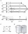
The Alp family of proteins. (A) Comparison of the four known members of the Alp protein family: the α, Rib, R28, and Alp2 proteins. S, signal peptide; N, nonrepeated N-terminal region; R, repeat region; C, C-terminal region. The number of amino acid residues in each region and the percent residue identity (bold numbers) between corresponding regions are indicated. Note that the designation Alp3 has been used as an alternative name (130) for the R28 protein (235). Each of the three proteins α, Rib, and R28 has 9 to 12 repeats with a length of 79 or 82 residues, as indicated. These repeats are identical within one protein but not between proteins, and the number of repeats varies among clinical isolates. The nonrepeated region of R28 contains a 195-residue subregion, designated RN, which does not fit into the alignment with Rib. This region is 99% identical to a type of repeat found in the N-terminal half of the Alp2 protein. Thus, the Alp2 protein contains two types of tandem repeat (designated RN and R), unlike the other members of the family. This figure is based on published sequences for the four proteins (78, 130, 165, 235, 261). The data for Alp2 are based on the sequence reported by Glaser et al. (78). (B) Schematic representation of the R28 protein. As indicated, R28 can be viewed as a chimera, derived from the two Alp family proteins α and Rib and the unrelated β protein (235). Predictions about the three-dimensional structure are given below the protein. The region that shows similarity to β corresponds to a region in β predicted to have an IgSF fold (12), and our analysis of the R28 region suggests that it has a similar structure. The repeats in the C-terminal part of R28 have been proposed to have a fold related to the IgSF fold (35). Modified from reference with permission of the publisher. (C) Immunological relationship among the four known members of the Alp protein family. Solid arrows indicate immunological cross-reactivity, and broken arrows indicate lack of cross-reactivity. For example, the α protein cross-reacts with Alp2 but not with Rib or R28. Most of the data summarized in the figure were obtained with rabbit antisera raised against purified proteins (132, 234-236), but the relationship between R28 and Alp2 was analyzed with an extract of strain D136C (NEM316), which expresses Alp2 (78, 234). (D) Laddering pattern in the α, Rib, and R28 proteins analyzed by SDS-PAGE. Purified preparations of the three proteins, and the control protein β (which is not a member of the Alp family), were subjected to SDS-PAGE under ordinary conditions (with boiling of samples at pH = 7) or after boiling of samples for 15 min at pH = 4. Under the latter conditions, the three Alp family proteins form a regularly laddered pattern due to hydrolysis of acid-sensitive Asp-Pro bonds in the repeats during sample preparation. Modified from reference with permission of the publisher.
FIG. 3.
FIG. 3.
Proteins in other bacterial species related to members of the Alp protein family. (A) Schematic representations of six Alp-related bacterial proteins. The bacterial species and the name of the protein are indicated to the left. For all proteins, the C-terminal part includes a region with long tandem repeats showing sequence similarity to the C-terminal repeat region of Alp family members. The lengths of the different repeats are indicated below each protein. The percentages indicate residue identity to the repeats of the Rib protein, the Alp family member to which these proteins show the greatest similarity. Note that the Esp protein of E. faecalis has a more complex structure than the other proteins and has three types of repeats, the tandem A and C repeats and the nontandem B repeats, of which the B and C repeats show similarity to the repeats of Rib and other Alp proteins. The complete sequence is not available for the Rib-like protein (Rlp) of L. fermentus. With one exception (BipA), these different proteins have an LPXTG motif in the C-terminal region, which may allow covalent anchoring to the cell wall. The BipA protein of the gram-negative bacterium B. pertussis is probably bound to the bacterium via an N-terminal hydrophobic region. The black boxes to the left represent signal peptides. See the text for details and references. (B) Alignment of sequences in a conserved region present within the repeats. Because these sequences are most similar to the corresponding sequence in Rib among different Alp proteins, the corresponding sequence in Rib is also included. Residues identical to those in Rib are shown in bold type, and the number of identities is indicated to the right. The consensus sequence represents residues present in at least five of the eight sequences.
FIG. 4.
FIG. 4.
Schematic representation of three S. agalactiae surface proteins with enzymatic and/or ligand-binding activity. (A) The ScpB protein, which was first identified as a C5a peptidase, has a Fn-binding region (13) that overlaps with the Ser-Asp-His catalytic triad required for enzyme activity (37). S, signal sequence. The C-terminal region (C) includes the putative wall-anchoring motif LPTTN. Modified from reference with permission. (B) The β protein has separate binding sites for human IgA-Fc (92, 107, 108, 214) and FH (5), as indicated. The XPZ region is a proline-rich and surface-exposed region with a highly periodic sequence (4). This region, which adopts the PPII helix structure (4), is not required for binding of FH (5). A region in the central part of the β protein is predicted to have an IgSF fold (12). The C-terminal region includes the putative wall-anchoring motif LPYTG. Modified from reference with permission of the publisher. (C) The FbsA protein is composed almost entirely of 16-residue repeats, which are responsible for the ability of the protein to bind Fg (219). The number of repeats varies among different bacterial strains. The C-terminal region includes the putative wall-anchoring motif LPKTG. Modified from reference with permission of the publisher.
FIG. 5.
FIG. 5.
Genetic structure of a chromosomal region, conserved between S. agalactiae and S. pyogenes, that includes genes for the surface proteins Scp and Lmb. The figure summarizes data from several studies (61, 73, 79, 197, 232, 251). The scp and lmb genes encode the C5a peptidase and the Lmb protein, respectively, while the function of ORF2 is unknown. These three genes are virtually identical in the two genomes, as indicated, while the surrounding chromosomal regions are unrelated (61, 73). In S. agalactiae, the three genes are located within a putative composite transposon that also includes several other genes and is bordered by IS elements, which are indicated in the figure but not drawn to scale (73). Many strains of S. agalactiae have the IS element IS1548 or the intron GBSi1 inserted between scpB and lmb. In S. pyogenes, the genes do not appear to be located within a transposon. In many (but not all) strains of S. pyogenes, the fba gene, which encodes the Fn-binding surface protein Fba, is located between scpA and lmb (61, 197, 250). In both species, the lmb and ORF2 genes are cotranscribed (61). In S. pyogenes the scpA gene is cotranscribed with fba, and both genes are controlled by the Mga protein, but a strong transcriptional attenuator is probably located between the two genes, as indicated (197).

Similar articles

Cited by

References

    1. Adderson, E. E., S. Takahashi, Y. Wang, J. Armstrong, D. V. Miller, and J. F. Bohnsack. 2003. Subtractive hybridization identifies a novel predicted protein mediating epithelial cell invasion by virulent serotype III group B Streptococcus agalactiae. Infect. Immun. 71:6857-6863. - PMC - PubMed
    1. Anderson, E. T., M. G. Wetherell, L. A. Winter, S. B. Olmsted, P. P. Cleary, and Y. V. Matsuka. 2002. Processing, stability, and kinetic parameters of C5a peptidase from Streptococcus pyogenes. Eur. J. Biochem. 269:4839-4851. - PubMed
    1. Areschoug, T., F. Carlsson, M. Stålhammar-Carlemalm, and G. Lindahl. 2004. Host-pathogen interactions in Streptococcus pyogenes infections, with special reference to puerperal fever and a comment on vaccine development. Vaccine 22:S9-S14. - PubMed
    1. Areschoug, T., S. Linse, M. Stålhammar-Carlemalm, L.-O. Hedén, and G. Lindahl. 2002. A proline-rich region with a highly periodic sequence in streptococcal β protein adopts the polyproline II structure and is exposed on the bacterial surface. J. Bacteriol. 184:6376-6383. - PMC - PubMed
    1. Areschoug, T., M. Stålhammar-Carlemalm, I. Karlsson, and G. Lindahl. 2002. Streptococcal β protein has separate binding sites for human factor H and IgA-Fc. J. Biol. Chem. 277:12642-12648. - PubMed

Publication types

MeSH terms

LinkOut - more resources