

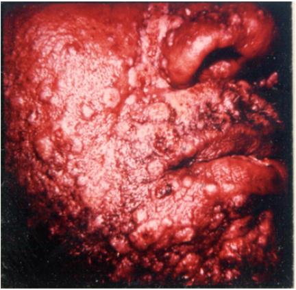
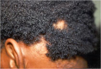
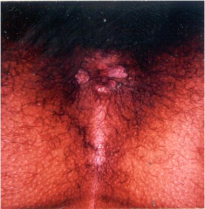
Secondary syphilitic lesions
- PMID: 15653827
- PMCID: PMC544174
- DOI: 10.1128/CMR.18.1.205-216.2005
Secondary syphilitic lesions
Abstract
An important theme that emerges from all early historical accounts is that in addition to the decreased virulence of Treponema pallidum, the incidence of secondary syphilis has decreased drastically over the past three centuries. Even in the early 20th century, most syphilologists were of the opinion that the disease had undergone changes in its manifestations and that they were dealing with an attenuated form of the spirochete. Such opinions were based primarily on the observations that violent cutaneous reactions and fatalities associated with the secondary stage had become extremely rare. The rate of primary and secondary syphilis in the United States increased in 2002 for the second consecutive year. After a decade-long decline that led to an all-time low in 2000, the recent trend is attributable, to a large extent, by a increase in reported syphilis cases among men, particularly homosexual and bisexual men having sex with men. The present review addresses the clinical and diagnostic criteria for the recognition of secondary syphilis, the clinical course and manifestations of the disease if allowed to proceed past the primary stage of disease in untreated individuals, and the treatment for this stage of the disease.
Figures


References
-
- Bayne, L. L., J. W. Schmidley, and D. S. Goodin. 1986. Acute syphilitic meningitis. Its occurrence after clinical and serologic cure of secondary syphilis with penicillin G. Arch. Neurol. 43:137-138. - PubMed
-
- Bauer, T. J., E. V. Price, and J. C. Cutler. 1952. Spinal fluid examinations among patients with primary or secondary syphilis. Am. J. Syph. 36:309-•••. - PubMed
-
- Baughn, R. E., M. C. McNeeley, J. L. Jorizzo, and D. M. Musher. 1986. Characterization of the antigenic determinants and host components in immune complexes from patients with secondary syphilis. J. Immunol. 136:1406-1414. - PubMed
-
- Baughn, R. E., and R. F. Schell. 2001. Immune responses to Treponema pallidum and Borrelia burgdorefi, p. 27.1-27.7. In R. R. Rich (ed.), Principles and practice of clinical immunology, 2nd ed. Mosby, St. Louis, Mo.
-
- Berry, C. D., T. M. Hooton, A. C. Collier, and S. A. Lukehart. 1987. Neurologic relapse after benzathine penicillin therapy for secondary syphilis in a patient with HIV infection. N. Engl. J. Med. 6:1587-1589. - PubMed
Publication types
MeSH terms
LinkOut - more resources
Full Text Sources
Miscellaneous
