Review
. 2005 Jan 15;118(Pt 2):283-90.
doi: 10.1242/jcs.01649.

The developmental cell biology of Trypanosoma brucei

Affiliations
Review

The developmental cell biology of Trypanosoma brucei

Keith R Matthews. J Cell Sci. .

Erratum in

  • J Cell Sci. 2005 May 1;118(Pt 9):2078

Abstract

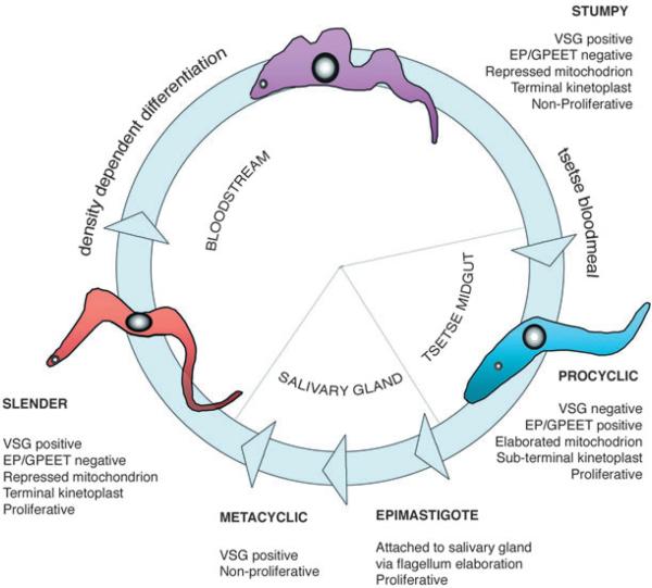
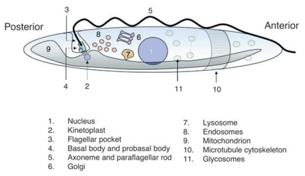
Trypanosoma brucei provides an excellent system for studies of many aspects of cell biology, including cell structure and morphology, organelle positioning, cell division and protein trafficking. However, the trypanosome has a complex life cycle in which it must adapt either to the mammalian bloodstream or to different compartments within the tsetse fly. These differentiation events require stage-specific changes to basic cell biological processes and reflect responses to environmental stimuli and programmed differentiation events that must occur within a single cell. The organization of cell structure is fundamental to the trypanosome throughout its life cycle. Modulations of the overall cell morphology and positioning of the specialized mitochondrial genome, flagellum and associated basal body provide the classical descriptions of the different life cycle stages of the parasite. The dependency relationships that govern these morphological changes are now beginning to be understood and their molecular basis identified. The overall picture emerging is of a highly organized cell in which the rules established for cell division and morphogenesis in organisms such as yeast and mammalian cells do not necessarily apply. Therefore, understanding the developmental cell biology of the African trypanosome is providing insight into both fundamentally conserved and fundamentally different aspects of the organization of the eukaryotic cell.


Figures

Fig. 1
Fig. 1. The life cycle of Trypanosoma brucei
Trypanosomes proliferate in the bloodstream of mammalian hosts as morphologically slender forms. These cells express the bloodstream-stage-specific VSG coat to evade the mammalian immune response. The kinetoplast (the mitochondrial genome of the parasite) is located at the posterior end of the cell and mitochondrial activity is relatively repressed. As parasite numbers increase in the bloodstream, differentiation to morphologically stumpy forms occurs. These are division-arrested forms pre-adapted for transmission to tsetse flies. Upon uptake in a tsetse bloodmeal, procyclic forms are generated, these being proliferative in the fly midgut. Procyclic forms express a surface coat distinct from that of bloodstream forms, the VSG being lost and replaced by a coat composed of EP and GPEET procyclins. The kinetoplast is also repositioned to a sub-terminal position. After establishment in the fly midgut, trypanosomes arrest in division and then migrate to the tsetse salivary gland, where they attach as epimastigote forms. These are proliferative and attached through elaboration of their flagellum. Eventually, these generate non-proliferative metacyclic forms, which have re-acquired a VSG coat in preparation for transmission to a new mammalian host. Arrowheads represent differentiation events in the trypanosome life cycle.
Fig. 2
Fig. 2. Trypanosome cell architecture
A simplified representation of the location of the major structural features of the trypanosome cell. A cutaway section towards the anterior of the cell shows the microtubule cytoskeleton underlying the cell membrane. For more detailed images of the trypanosome cell, the reader is referred to recent articles (Grunfelder et al., 2003; Overath and Engstler, 2004; Vaughan and Gull, 2003).
Fig. 3
Fig. 3. The tripartite attachment complex
(A) An electron micrograph of the region of the flagellar pocket (labelled F.P). The basal body (BB) is shown connected to the kinetoplast through a series of unilateral filaments, which link the kinetoplast to the inner mitochondrial membrane (these are indicated by the small brackets). The exclusion zone filaments (indicated by a large bracket) link the mitochondrial outer membrane and the basal body. The inset shows a schematic representation of the region of the trypanosome cell shown in the electron micrograph. (B) A representation of the flagellar/basal bodies/kinetoplast region and its replication during the trypanosome cell cycle. The left-hand image is the organization of the tripartite attachment complex in G1 phase; the middle image represents S phase, when kinetoplast replication is underway, and when the probasal body has matured and two new probasal bodies have formed. The right-hand image shows segregation of the basal bodies and concomitant kinetoplast DNA segregation. Figure adapted with kind permission from K. Gull (University of Oxford, UK) and The American Society for Cell Biology (Ogbadoyi et al., 2003).
Fig. 4
Fig. 4. The kinetoplast changes position during the trypanosome life cycle
(A) The relative position of the kinetoplast in trypomastigote bloodstream and procyclic forms and in epimastigote forms with respect to the nucleus and posterior end of the trypanosome. (B) A phase contrast image of cells undergoing differentiation between bloodstream stumpy forms and procyclic forms. The cells have been counter-stained with DAPI to reveal the position of the nucleus (N) and kinetoplast (K). In the three cells shown, the kinetoplast is progressively repositioned, being earliest in this process in the top cell and latest in the bottom cell. Panel B is reproduced from Matthews et al. (Matthews et al., 1995). Bar, 20 μm.
Fig. 5
Fig. 5. The morphology of procyclic-form trypanosomes induced by ectopic overexpression of TbZFP2
(A) A phase contrast image of procyclic forms, one of which shows a greatly extended posterior end (nozzle). (B) A composite of the DNA of the trypanosome cell (labelled with DAPI and pseudocoloured blue; each cell contains a central nucleus and posterior kinetoplast) and staining with an antibody (YL1/2; Kilmartin et al., 1982) against tyrosinated α-tubulin, which labels dynamic microtubules in the trypanosome cytoskeleton (pseudocoloured green). Note that the posterior end of each cell shows staining, as does the newly growing daughter flagellum (labelled F) and the basal bodies (labelled in two of the cells with an asterisk). The nozzle cell shows an extended region of staining at the posterior end of the cell, indicating microtubule extension in this region. Bar, 8 μm.

Similar articles

Cited by

References

    1. Allen CL, Goulding D, Field MC. Clathrin-mediated endocytosis is essential in Trypanosoma brucei. EMBO J. 2003;22:4991–5002. - PMC - PubMed
    1. Amiguet-Vercher A, Perez-Morga D, Pays A, Poelvoorde P, van Xong H, Tebabi P, Vanhamme L, Pays E. Loss of the mono-allelic control of the VSG expression sites during the development of Trypanosoma brucei in the bloodstream. Mol. Microbiol. 2004;51:1577–1588. - PubMed
    1. Bastin P, Sherwin T, Gull K. Paraflagellar rod is vital for trypanosome motility. Nature. 1998;391:548. - PubMed
    1. Bochud-Allemann N, Schneider A. Mitochondrial substrate level phosphorylation is essential for growth of procyclic Trypanosoma brucei. J. Biol. Chem. 2002;277:32849–32854. - PubMed
    1. Briggs LJ, McKean PG, Baines A, Moreira-Leite F, Davidge J, Vaughan S, Gull K. The flagella connector of Trypanosoma brucei: an unusual mobile transmembrane junction. J. Cell Sci. 2004;117:1641–1651. - PubMed

Publication types

MeSH terms