. 2005 Mar 25;280(12):11552-9.
doi: 10.1074/jbc.M410033200. Epub 2005 Jan 14.

R1, a novel repressor of the human monoamine oxidase A

Affiliations

R1, a novel repressor of the human monoamine oxidase A

Kevin Chen et al. J Biol Chem. .

Abstract

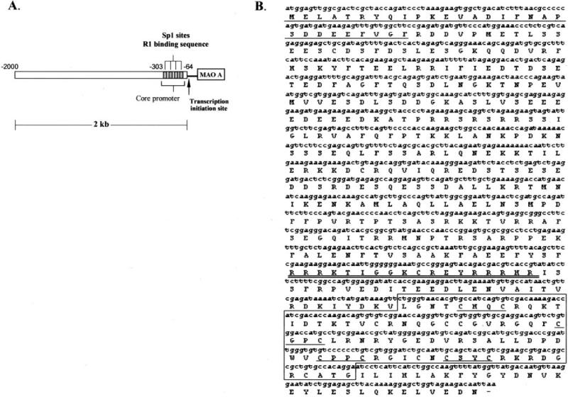
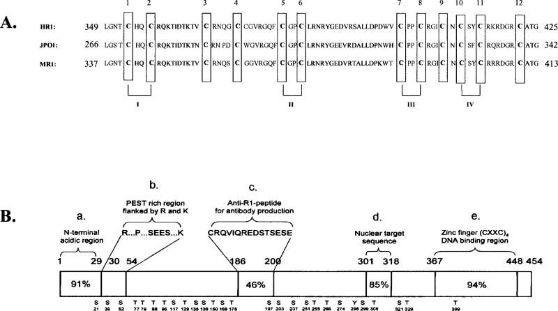
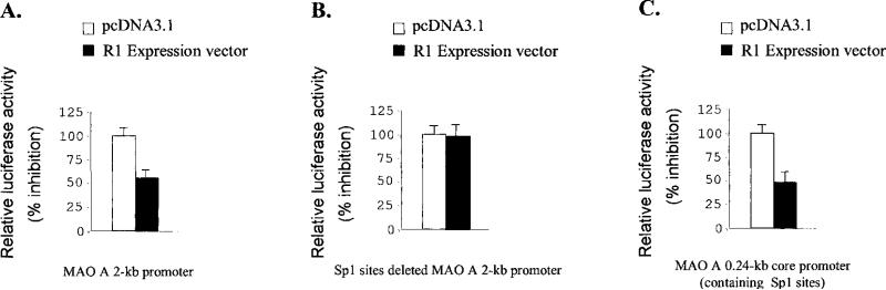
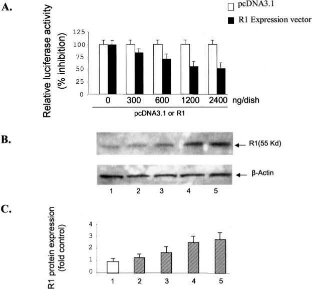
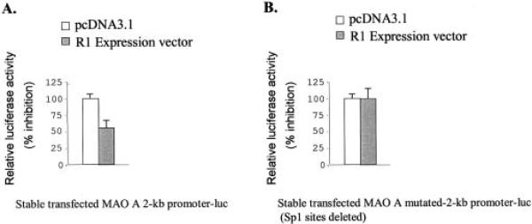
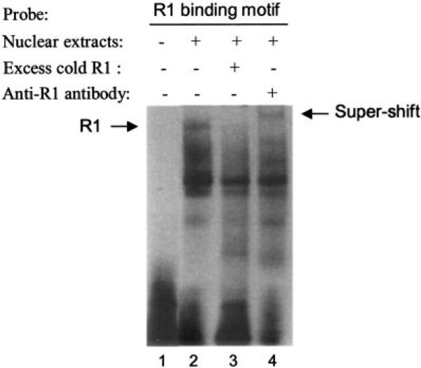
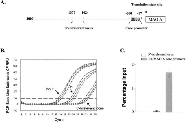
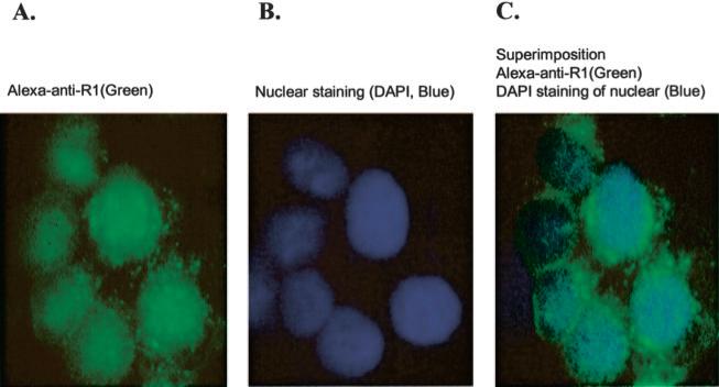
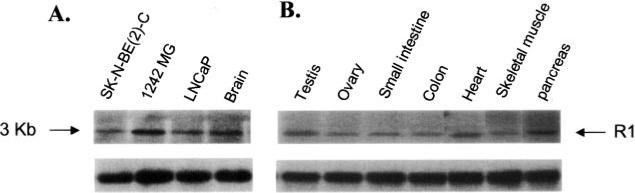
Monoamine oxidase catalyzes the oxidative deamination of a number of neurotransmitters. A deficiency in monoamine oxidase A results in aggressive behavior in both humans and mice. Studies on the regulation of monoamine oxidase A gene expression have shown that the Sp1 family is important for monoamine oxidase A expression. To search for novel transcription factors, the sequences of three Sp1 sites in the monoamine oxidase A core promoter were used in the yeast one-hybrid system to screen a human cDNA library. A novel repressor, R1 (RAM2), has been cloned. The R1 cDNA encodes a protein with 454 amino acids and an open reading frame at the 5'-end. The transfection of R1 in a human neuroblastoma cell line, SK-N-BE (2)-C, inhibited the monoamine oxidase A promoter and enzymatic activity. The degree of inhibition of monoamine oxidase A by R1 correlated with the level of R1 protein expression. R1 was also found to repress monoamine oxidase A promoter activity within a natural chromatin environment. A gel-shift assay indicated that the endogenous R1 protein in SK-N-BE (2)-C cells interacted with the R1 binding sequence. R1 also bound directly to the natural monoamine oxidase A promoter in vivo as shown by chromatin immunoprecipitation assay. Immunocytochemical analysis showed that R1 was expressed in both cytosol and nucleus, which suggested a role for R1 in transcriptional regulation. Northern blot analysis revealed the presence of endogenous R1 mRNA in human brain and peripheral tissues. Taken together, this study shows that R1 is a novel repressor that inhibits monoamine oxidase A gene expression.


Figures

Fig. 1
Fig. 1
A, the human MAO A promoter 2-kb region showing the location of R1 binding sequence (Sp1 sites) used as a probe for yeast one-hybrid system to screen the novel R1 protein. The core promoter region (−303 to −64 bp) and the transcription initiation site were indicated. B, the human R1 cDNA sequence and deduced amino acid sequence. Several important protein domains were indicated, the N-terminal acidic region (amino acids 1–29) was underlined; the nuclear targeting sequence between amino acids 301 and 318 was double underlined, and the 77 amino acid residues (amino acids 349–425) of the C-terminal including DNA-binding domains were boxed. The 4 CXXC zinc finger putative DNA binding domains were underlined.
Fig. 2
Fig. 2. Potential functional domains in human R1 repressor protein predicted by Prosite analysis
A, an alignment of the 77 amino acid residues of the C-terminal of human R1 protein (HR1), JP01, and mouse repressor 1 (MR1). 4 CXXC zinc finger putative DNA binding domains were indicated by I, II, III, and IV at the bottom. The conserved cysteine residues are marked and boxed and numbered from 1 to 12 at the top. B, several functional domains with amino acid location were indicated at the top. The percent identity between human and mouse R1 protein was obtained by blast analysis. Bottom, potential phosphorylation sites conserved in human and mouse R1. Serine, S; threonine, T; tyrosine, Y. Numbers denotes the amino acid residues in R1 protein sequence.
Fig. 3
Fig. 3. The effect of repressor R1 on MAO A catalytic activity in SK-N-BE (2)-C cells
A, the repressor R1 construct driven by CMV promoter or pcDNA3.1 vector were transfected into SK-N-BE (2)-C cells. 24 h later, cells were replated into 5-cm dishes, and Geneticin (G418; 600 μg/ml) was added. Resistant clones have been isolated after 6 days and cultured under continuous G418 selection. MAO A enzymatic activity was determined in the stable cells from five separate clones to evaluate the function of R1. B, the level of stably expressed R1 was evaluated by Western blot using anti-R1 antibody. C, the intensities of bands were quantified by PhosphorImager. Values were normalized to actin levels on the corresponding reprobed filters and then expressed as the -fold of control, in which the control was taken as 1. Data represented the mean ± S.E. of three independent experiments.
Fig. 4
Fig. 4. The effect of repressor R1 on MAO A promoter activity in SK-N-BE (2)-C cells
MAO A 2-kb promoter-luciferase construct (A), Sp1 sites deleted MAO A 2-kb promoter-luciferase construct (B), or MAO A core promoter containing Sp1 sites (0.24-kb)-luciferase construct (C) were cotransfected with R1 expression vector into SK-N-BE (2)-C cells. After 48 h, the luciferase activity was determined. All data are presented as the mean ± S.E. of at least three independent experiments. The controls were transfected with pcDNA3.l.
Fig. 5
Fig. 5. The degree of repression of R1 on MAO A promoter activity correlated with the level of R1 expression
Different amounts of R1 or pcDNA3.1 vector (0, 300, 600, 1200, and 2400 ng/dish) were cotransfected with MAO A 2-kb-luc into SK-N-BE (2)-C cells for 2 days and either the luciferase activity was determined (A), or nuclear proteins were extracted and analyzed by Western blot using anti-R1 antibody (B). The relative intensity of each R1 band was quantified by PhosphorImager (C). Values were normalized to actin levels on the corresponding reprobed filters and then expressed as the -fold of control in which the control was taken as 1. Data represented the mean ± S.E. of three independent experiments.
Fig. 6
Fig. 6. The repressor R1 inhibited MAO A promoter activity in the context of a chromatinized template
SK-N-BE (2)-C cells were stably transfected with the wild-type or Sp1 sites deleted MAO A 2-kb-luc construct to generate the cell line with a MAO A 2-kb-luc stably integrated into the genome. After Geneticin (G418; 600 μg/ml) selection, resistant clones were isolated and cultured under continuous G418 selection. Then R1 expression vector or pcDNA3.1 (control) was transfected into cells with stably expressed wild-type (A) or Sp1 sites deleted MAO A 2-kb-luc (B). After 48 h, the luciferase activity was determined. All data are presented as the mean ± S.E. of at least three independent experiments.
Fig. 7
Fig. 7. The novel repressor protein R1 was present in nuclear extracts of SK-N-BE (2)-C cells and was bound to R1 binding sequence
The gel-shift assay was performed with 20 μg of nuclear extracts from SK-N-BE (2)-C cells (lanes 2, 3, and 4) and the 32P-labeled R1 binding sequence (Sp1 sites), which was used in the yeast one-hybrid system to fish out R1. Excess cold R1 binding motif probe (lane 3)or anti-R1 antibody (lane 4) was added as indicated. An arrow indicates the R1-DNA complex. The super-shifted complex is also indicated.
Fig. 8
Fig. 8. R1 interacted with the endogenous MAO A promoter directly in vivo
The occupation of R1 on the MAO A core promoter or 5′-irrelevant region was determined by ChIP assay combined with quantitative real time PCR using SK-N-BE (2) C cells. A, a schematic representation of the MAO A promoter. Real time PCR-targeted regions containing core promoter and Sp1 sites (from −360 to −17 bp) and 5′-irrelevant loci (from −1377 to −1024 bp for negative control) were indicated. B, representative R1 ChIP/quantitative real time PCR amplification plots (triplicates). The ChIP/quantitative real time PCR amplification was performed for cross-linked inputs, R1-associated MAO A core promoter, or 5′-irrelevant loci in SK-N-BE (2) C cells. The nuclear protein-DNA complex was immunoprecipitated by anti-R1 antibody and was quantitatively analyzed by real time PCR. C, analysis of association of R1 with MAO A core promoter or 5′-irrelevant locus. The relative differences between input sample and R1 or negative control were determined using the ΔCT method (see “Materials and Methods”). These values were presented as percent input in which the DNA cross-linked input sample was taken as 100%. Data were the mean ± S.D. from triplicate samples of three independent experiments.
Fig. 9
Fig. 9. Repressor R1 was located in both nucleus and cytosol of human neuroblastoma SK-N-BE (2)-C cells
Immunofluorescence was performed using anti-R1 antibody. SK-N-BE (2)-C cells were plated on coverslips the day before experiment. Then the cells were fixed and incubated with rabbit anti-R1 antibody, followed with fluorescein-conjugated anti-rabbit secondary antibody (A, green). The stained slides were mounted in the presence of 4, 6-diamino-2-phenylindole (DAPI) for nuclear staining (B, blue) and examined under fluorescence microscope. C, the merge of A and B.
Fig. 10
Fig. 10. The presence of endogenous R1 mRNA (3 kb) in human brain, peripheral tissues, and cell lines as shown by Northern blot analysis
A, 10 μg of total RNA from SK-N-BE (2)-C (neuroblastoma), 1242-MG (astrocytoma), LNCaP (prostate cancer), and human brain samples. B, 2 μg of poly(A)+ RNA from human peripheral tissues as indicated were loaded on each gel and then transferred to membranes. A 500-bp EcoRI fragment R1-specific probe was labeled with 32P and hybridized to the membranes overnight at 42 °C. Blots were exposed for 16 h. Bottom blots, the same blots were hybridized with glyceraldehyde-3-phosphate dehydrogenase positive control probe and hybridized and washed under the same condition. The film was exposed for4hto demonstrate that the same amount of RNA was loaded on each lane.

Similar articles

Cited by

References

    1. Shih JC. Neuropsychopharmacology. 1991;4:1–7. - PubMed
    1. Thorpe LW, Westlund KN, Kochersperger LM, Abell CW, Denney RM. J. Histochem. Cytochem. 1987;35:23–32. - PubMed
    1. Johnston JP. Biochem. Pharmacol. 1968;17:1285–1297. - PubMed
    1. Knoll J, Magyar K. Adv. Biochem. Psychopharmacol. 1972;5:393–408. - PubMed
    1. Bach AW, Lan NC, Johnson DL, Abell CW, Bembenek ME, Kwan SW, Seeburg PH, Shih JC. Proc. Natl. Acad. Sci. U. S. A. 1988;85:4934–4938. - PMC - PubMed

Publication types

LinkOut - more resources