. 2005 Jan 25;44(3):971-8.
doi: 10.1021/bi048208a.

Mass spectrometric identification of lysines involved in the interaction of human replication protein a with single-stranded DNA

Affiliations

Mass spectrometric identification of lysines involved in the interaction of human replication protein a with single-stranded DNA

Steven M Shell et al. Biochemistry. .

Abstract

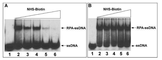
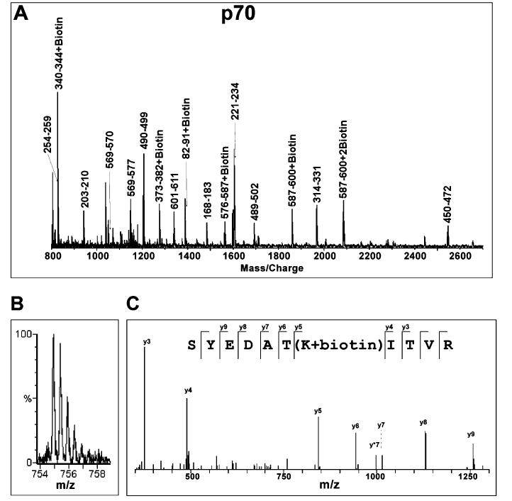
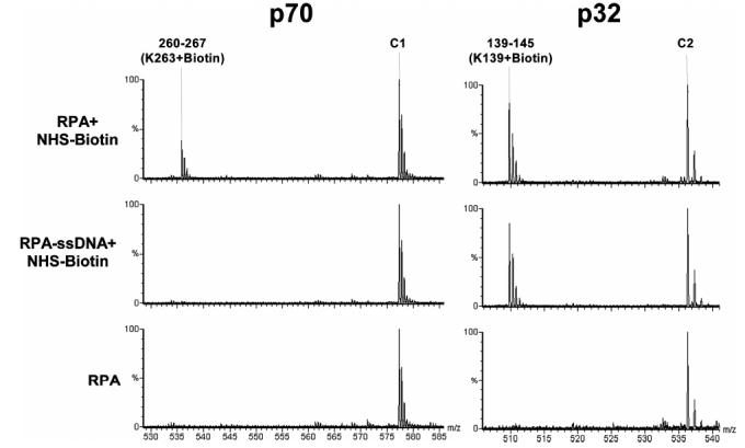
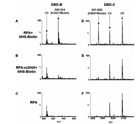
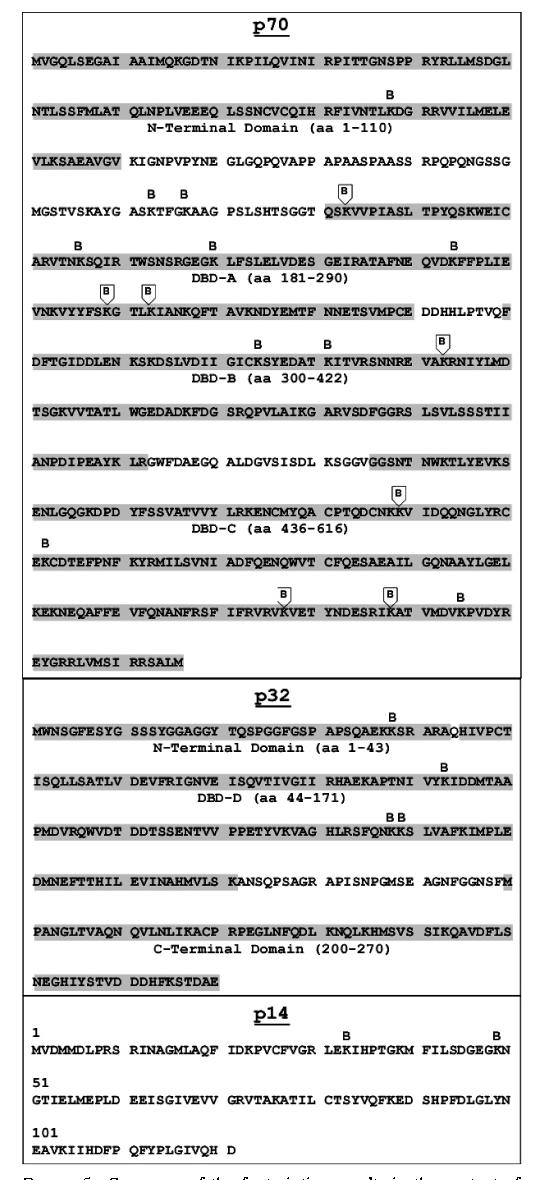
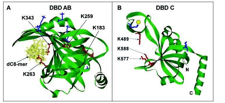
Human replication protein A (hRPA), a heterotrimeric single-stranded DNA (ssDNA) binding protein, is required for many cellular pathways including DNA damage repair, recombination, and replication as well as the ATR-mediated DNA damage response. While extensive effort has been devoted to understanding the structural relationships between RPA and ssDNA, information is currently limited to the RPA domains, the trimerization core, and a partial cocrystal structure. In this work, we employed a mass spectrometric protein footprinting method of single amino acid resolution to investigate the interactions of the entire heterotrimeric hRPA with ssDNA. In particular, we monitored surface accessibility of RPA lysines with NHS-biotin modification in the contexts of the free protein and the nucleoprotein complex. Our results not only indicated excellent agreement with the available crystal structure data for RPA70 DBD-AB-ssDNA complex but also revealed new protein contacts in the nucleoprotein complex. In addition to two residues, K263 and K343 of p70, previously identified by cocrystallography as direct DNA contacts, we observed protection of five additional lysines (K183, K259, K489, K577, and K588 of p70) upon ssDNA binding to RPA. Three residues, K489, K577, and K588, are located in ssDNA binding domain C and are likely to establish the direct contacts with cognate DNA. In contrast, no ssDNA-contacting lysines were identified in DBD-D. In addition, two lysines, K183 and K259, are positioned outside the putative ssDNA binding cleft. We propose that the protection of these lysines could result from the RPA interdomain structural reorganization induced by ssDNA binding.


Figures

F<sc>igure</sc> 1:
Figure 1:
Effects of biotinylation on RPA-ssDNA binding. Lanes: 1, free ssDNA; 2-6, ssDNA bound by RPA pre- or posttreated with 0, 50, 100, 200, and 400 μM NHS-biotin. Panel A: Addition of increasing amounts of NHS-biotin prior to addition of ssDNA blocked critical lysine residues for ssDNA interaction and thus abolished binding affinity. Panel B: Addition of ssDNA prior to NHS-biotin, however, shielded critical lysine residues from modification and showed little effect of biotin on the formed RPA-ssDNA complex.
F<sc>igure</sc> 2:
Figure 2:
Mass spectrometric analysis of biotinylation of RPA protein. (A) A typical MALDI-TOF spectrum of tryptic digestion of biotinylated RPA70. Monoisotopic resolution for all of the peptide peaks was obtained, allowing unequivocal assignment of singly charged unmodified and biotinylated peptide fragments. (B) A typical Q-TOF spectrum of the doubly charged biotinylated p70 peptide fragment. The molecular mass of this ion in the form of m/z (mass per charge) corresponds to peptide fragment 325-335 + biotin. (C) MS/MS analysis of the parent ion (m/z) 754.91) shown in (B) confirms that lysine 331 is biotinylated. The y ion series were derived from internal fragmentation of the peptide bonds, providing sequence information read from the C-terminus (left) to the N-terminus (right). The mass increment between y4 and y5 ions corresponds to a biotinylated lysine while the masses of the remaining y ions correspond perfectly to the amino acid sequence of the fragment.
F<sc>igure</sc> 3:
Figure 3:
Identification of lysine residues protected from modification by nucleic acid. The typical Q-TOF data illustrate the protection of a lysine residue in p70 versus an unprotected lysine in p32. The peak corresponding to peptide fragment 260-267 + biotin in p70 is an example of protection from modification by binding of ssDNA. When ssDNA is not present, K263 is readily modified by NHS-biotin. In the presence of ssDNA, however, the modification peak disappears. In contrast, K139 in p32 is a lysine residue not protected by ssDNA binding. A modification peak appears upon treatment with NHS-biotin and persists following addition of ssDNA. Each multiply charged peptide resulted in clearly resolved peak clusters, indicating monoisotopic resolution; unmodified peaks C1 and C2 serve as controls.
F<sc>igure</sc> 4:
Figure 4:
MALDI-TOF analysis of protection of lysine residues. The peptide peak containing modified K343 of DBD-B (A) is absent in the presence of the dT30mer (B). Similarly, K588 is located in DBD-C and is biotinylated in the absence of ssDNA (D) but is protected from modification by direct contact with the dT30mer (E). Panels C and F show the spectra of RPA without treatment of biotin. The unmodified peaks C3, C4, and C5 all serve as controls.
F<sc>igure</sc> 5:
Figure 5:
Summary of the footprinting results in the context of the RPA sequence. Biotinylation sites in the three subunits of RPA are indicated in the primary amino acid sequence either as protected (boxed B) or as unprotected residues (unboxed B). The locations of domain structures are indicated by the shaded sequence with the name and amino acid numbering of the structure.
F<sc>igure</sc> 6:
Figure 6:
Structural exhibition of modified lysines in DBD-AB and DBD-C. (A) Structure of the DBD-AB-ssDNA complex (13). Lysine residues in the structures are presented in stick representation. K263 and K343 are found in the binding clefts of DBD-A and DBD-B, respectively, and are protected from modification by direct contact with the dC8mer. Lysine residues K183 and K259 are not in direct contact with the dC8mer but are protected from modification when ssDNA is present. (B) Structure of DBD-C (21). Lysine residues K489, K577, and K588 are located in the binding cleft of DBD-C and are protected from biotin modification when ssDNA is present, indicating direct contact with ssDNA as it transverses the binding cleft. Biotin-modified lysine residues without protection are shown in blue while the lysine residues protected from modification in the presence of ssDNA are shown in red in each structure.

Similar articles

Cited by

References

    1. Wold MS. Replication Protein A: A Heterotrimeric, Single-Stranded DNA-Binding Protein Required for Eukaryotic DNA Metabolism. Annu. ReV. Biochem. 1997;66:61–92. - PubMed
    1. Iftode C, Daniely Y, Borowiec JA. Replication Protein A (RPA): The Eukaryotic SSB. Crit. ReV. Biochem. Mol. Biol. 1999;34:141–180. - PubMed
    1. Stigger E, Dean FB, Hurwitz J, Lee SH. Reconstitution of Functional Human Single-Stranded DNA-Binding Protein from Individual Subunits Expressed by Recombinant Baculoviruses. Proc. Natl. Acad. Sci. U.S.A. 1994;91:579–583. - PMC - PubMed
    1. Henricksen LA, Umbricht CB, Wold MS. Recombinant Replication Protein A: Expression, Complex Formation, and Functional Characterization. J. Biol. Chem. 1994;269:11121–11132. - PubMed
    1. Burns JL, Daniely Y, Borowiec JA. Crit. ReV. Biochem. Mol. Biol. 1999;34:141–180. - PubMed

Publication types