. 2005 Feb;139(2):225-35.
doi: 10.1111/j.1365-2249.2004.02684.x.

Mycophenolate mofetil and roscovitine decrease cyclin expression and increase p27(kip1) expression in anti Thy1 mesangial proliferative nephritis

Affiliations

Mycophenolate mofetil and roscovitine decrease cyclin expression and increase p27(kip1) expression in anti Thy1 mesangial proliferative nephritis

M Chiara et al. Clin Exp Immunol. 2005 Feb.

Abstract

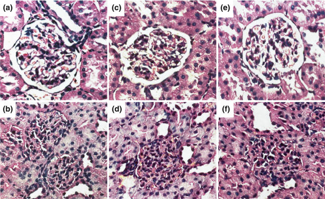
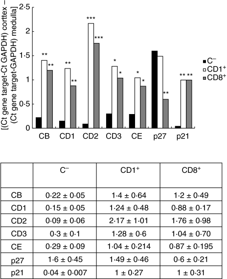
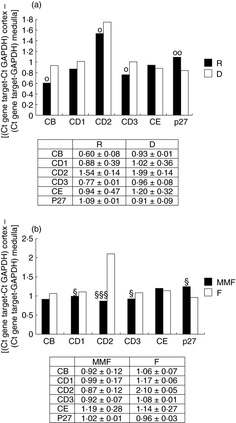
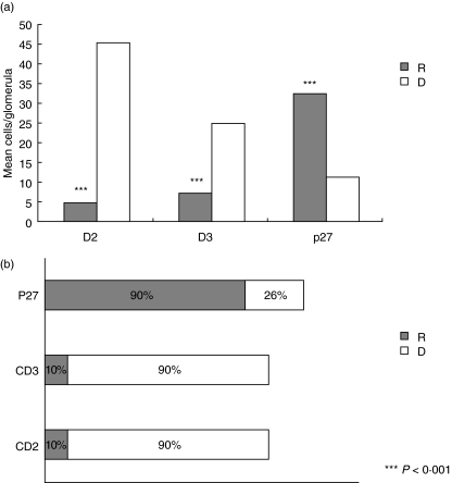
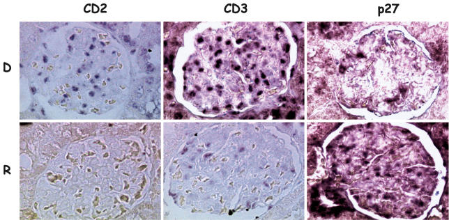
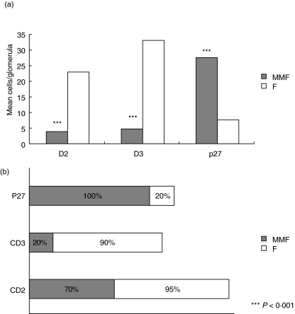
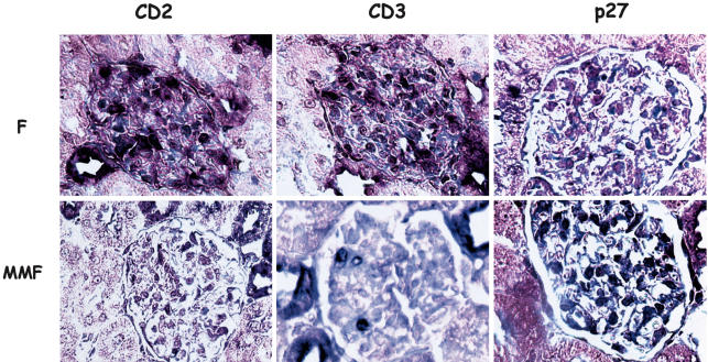
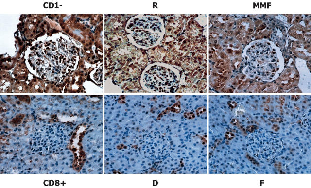
The response of mesangial cells to a phlogistic challenge includes cell proliferation and mesangial matrix expansion. Cell proliferation is a highly regulated process which includes enhancing factors such as cyclins, cyclin dependent kinases, and inhibitory proteins, such as p27(kip1). The aim of the study was to evaluate the effects of Mycophenolate mofetil (MMF), and roscovitine (R), on the cell cycle regulatory system when administered in the florid phase of the experimental model of mesangial proliferative nephritis induced by the anti Thy-1 antigen monoclonal antibody. Three days after nephritis induction, different groups were given MMF and R. Rats treated with MMF or R showed a slight decrease in mesangial proliferation and matrix expansion. Samples of cortical tissue were tested by 'real time' RT-PCR in order to study gene expression of cyclins B, D1, D2, D3, E, and the cyclin inhibitor p27(kip1). Localization of mRNA was evaluated by in situ hybridization. Real time RT-PCR analysis showed a significant decrease in cyclins B, D1, D2, and D3 in rats treated with either MMF or R as compared to controls. Both MMF and R treatment induced a significant increase in p27(kip1) mRNA expression. In situ hybridization showed a mesangial-endothelial expression pattern in glomeruli. The number of labelled cells per glomerulus, the number of positive glomeruli in each examined slide as well as cyclin D2 and D3 signal intensity was significantly lower in rats treated with MMF or R as compared to controls, whereas MMF or R treatment up-regulated p27(kip1) mRNA expression. Immunohistochemical evaluation of p27(kip1) aimed to examine the influence of MMF or R on protein expression confirmed up-regulation.


Figures

Fig. 1
Fig. 1
Morphological analyses showing glomerular cell proliferation and matrix expansion 8 days after glomerulonephritis induction (a) normal rats, (b) nephritic rats (CD8+) sacrificed 8 days after nephritis induction: an extensive mesangial expansion obliterating the urinary space can be observed. Compared to controls, (d) DMSO and (f) buffered saline, rats treated with MMF (e) or Roscovitine (c) showed improvement in histological changes.
Fig. 2
Fig. 2
Cyclin and p27 gene expression in cortical renal tissue of nephritic rats treated with the anti Thy 1 monoclonal antibody.Compared to normal rats, the cortical tissue of nephritic animals already showed increased gene expression for D-type cyclins and for cyclin B one day after disease induction (phase I, CD1+ group). This up-regulation was maintained throughout the proliferative phase (until the eighth day, phase II, CD8+ rats). Gene expression of the CDK inhibitor p27, which was found to be intensively positive in CD1+ nephritic rats as well as in normal ones, considerably decreased during mesangial cell proliferation (phase II, CD8+ rats). All gene expression data are expressed as [(gene target in cortex – GAPDH in cortex) – (gene target in medulla -GAPDH in medulla)]. Levels of significance refer to CD1+ and CD8+ compared to C-: ***P < 0·001; **P < 0·01; *P < 0·05.
Fig. 3
Fig. 3
Cyclin and p27 gene expression in cortical renal tissue of nephritic rats treated with mycophenolate mofetil and roscovitine. (a) Plot of gene expression data of the rats treated with roscovitine (R) and roscovitine vehicle (DMSO, D). (b) Plot of gene expression data of the rats treated with MMF and MMF vehicle (buffered saline, F). All gene expression data are expressed as [(gene target in cortex – GAPDH in cortex) – (gene target in medulla -GAPDH in medulla)]. Levels of significance refer to R compared to D: °°P < 0·01; °P < 0·05; MMF compared to F: §§§P < 0·001; §P < 0·05.
Fig. 4
Fig. 4
In situ hybridization: quantification of labelling after roscovitine treatment. (a) plot of the number of labelled cells per glomerulus (b) plot of the percentage of labelled glomeruli per section.Labelling was evaluated in at least 4 rats per treatment, in 2 sections per rat, and in at least 50 glomeruli per section. R, roscovitine; D, DMSO (roscovitine vehicle). Levels of significance refer to R compared to D: ***P < 0·001.
Fig. 5
Fig. 5
In situ hybridization in roscovitine treated rats. Decrease in signal intensity for group D cyclins and increase in p27 gene expression in roscovitine (R) treated rats compared to controls receiving DMSO (D).
Fig. 6
Fig. 6
In situ hybridization: quantification of labelling after mycophenolate mofetil treatment. (a) plot of the number of labelled cells per glomerulus. (b) plot of the percentage of labelled glomeruli per section.Labelling was evaluated in at least 4 rats per treatment, in 2 sections per rat, and in at least 50 glomeruli per section.MMF = mycophenolate mofetilF = buffered saline (MMF vehicle) Levels of significance refer to MMF compared to F: ***P < 0·001.
Fig. 7
Fig. 7
In situ hybridization in mycophenolate mofetil treated rats. Decrease in signal intensity for group D cyclins and increase in p27 gene expression in MMF-treated rats (MMF) compared to controls receiving buffered saline (F).
Fig. 8
Fig. 8
p27 expression. p27kip1 in untreated rats (CD1-) was found to be diffusely expressed both in mesangial and in epithelial cells, as per examination by immunohistochemical techniques, while CD1+ rats presented a decrease in labelled glomeruli (not shown). p27kip1 expression was completely abrogated in nephritic rats (CD8+), where tissue sections showed clear glomerular hypercellularity with a reduction in urinary space. Compared to controls (DMSO, D and saline, F) both Roscovitine (R) and Mycophenolate mofetil MMF treatment induced a significant up-regulation of p27kip1 expression.

Similar articles

Cited by

References

    1. Ooi BS, Cohen DJ, Veis JH. Biology of the mesangial cell in glomerulonephritis- role of cytokines. Proc Soc Exp Biol Medical. 1996;213:230–7. - PubMed
    1. Couser WG. Pathogenesis of glomerular damage in glomerulonephritis. Nephrol Dial Transplant. 1998;13:10–5. - PubMed
    1. Pines J. Cyclin and cyclin-dependent kinases: a biochemical view. Biochem J. 1995;308:697–711. - PMC - PubMed
    1. Sherr CJ. Mammalian G1 cyclins. Cell. 1993;73:1059–65. - PubMed
    1. Weiberg RA. The retinoblastoma protein and cell cycle control. Cell. 1995;81:323–30. - PubMed

MeSH terms