

Autoantibodies directed against the protease inhibitor calpastatin in psoriasis
- PMID: 15654835
- PMCID: PMC1809283
- DOI: 10.1111/j.1365-2249.2005.02701.x
Autoantibodies directed against the protease inhibitor calpastatin in psoriasis
Abstract
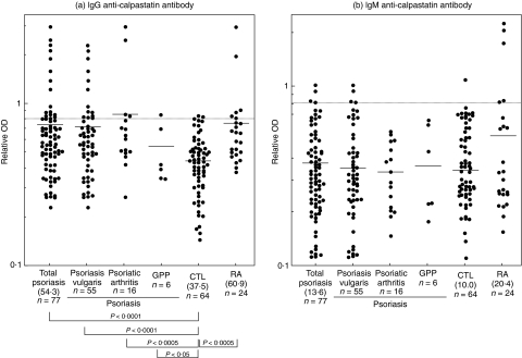
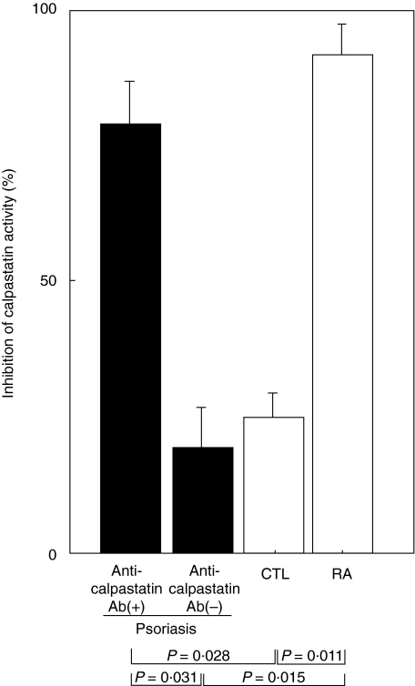
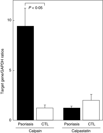
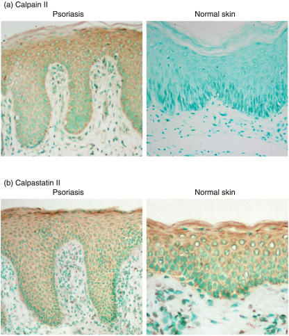
Psoriasis is believed to be a T cell-mediated autoimmune disease, but also exhibits autoantibody production. Calpastatin is an endogenous inhibitor of calpain, a ubiquitous protease that regulates inflammatory processes. Anti-calpastatin autoantibody was first identified as an autoantibody specific to rheumatoid arthritis, but has been also detected in other autoimmune diseases. In this study, we examined the presence and levels of anti-calpastatin antibody in 77 psoriasis patients by enzyme-linked immunosorbent assay. Compared with normal controls, psoriasis patients exhibited significantly elevated IgG anti-calpastatin antibody levels that were similar to those found in rheumatoid arthritis patients. Remarkably, IgG anti-calpastatin autoantibody in sera from psoriasis patients inhibited calpastatin activity. Calpain II expression was up-regulated in psoriasis skin lesions compared with normal skin while calpastatin expression was normal. The results of this study reveal the presence of anti-calpastatin autoantibody in psoriasis.
Figures
Similar articles
-
Autoantibodies to calpastatin (an endogenous inhibitor for calcium-dependent neutral protease, calpain) in systemic rheumatic diseases.Proc Natl Acad Sci U S A. 1995 Aug 1;92(16):7267-71. doi: 10.1073/pnas.92.16.7267. Proc Natl Acad Sci U S A. 1995. PMID: 7638179 Free PMC article.
-
High diagnostic value of anticalpastatin autoantibodies in rheumatoid arthritis detected by ELISA using human erythrocyte calpastatin as antigen.J Rheumatol. 2004 Jan;31(1):17-22. J Rheumatol. 2004. PMID: 14705213
-
[Antigenic epitopes recognized by autoantibodies to calpastatin in patients with rheumatoid arthritis and their clinical significance].Ryumachi. 1997 Jun;37(3):458-66. Ryumachi. 1997. PMID: 9256029 Clinical Trial. Japanese.
-
[Anti-calpastatin antibody as a serological marker for the diagnosis of rheumatoid arthritis].Nihon Rinsho. 2005 Jan;63 Suppl 1:344-50. Nihon Rinsho. 2005. PMID: 15799375 Review. Japanese. No abstract available.
-
[RA-related autoantibodies (anti-calpastatin, anti-gp130-RAPS, and anti-FRP antibodies)].Nihon Rinsho. 2005 Jul;63 Suppl 7:449-52. Nihon Rinsho. 2005. PMID: 16111298 Review. Japanese. No abstract available.
Cited by
-
Changes in Proteome of Fibroblasts Isolated from Psoriatic Skin Lesions.Int J Mol Sci. 2020 Jul 28;21(15):5363. doi: 10.3390/ijms21155363. Int J Mol Sci. 2020. PMID: 32731552 Free PMC article.
-
Differentially expressed proteins in the skin mucus of Atlantic cod (Gadus morhua) upon natural infection with Vibrio anguillarum.BMC Vet Res. 2013 May 14;9:103. doi: 10.1186/1746-6148-9-103. BMC Vet Res. 2013. PMID: 23672475 Free PMC article.
-
Prevalence of autoantibodies in patients of psoriasis.J Clin Lab Anal. 2010;24(1):44-8. doi: 10.1002/jcla.20365. J Clin Lab Anal. 2010. PMID: 20087953 Free PMC article.
-
The Future Landscape of Endothelial Cells Research in Psoriasis: Bibliometric Analysis and Literature Review.Clin Cosmet Investig Dermatol. 2023 Oct 30;16:3107-3120. doi: 10.2147/CCID.S435085. eCollection 2023. Clin Cosmet Investig Dermatol. 2023. PMID: 37927385 Free PMC article.
-
CAPN6 in disease: An emerging therapeutic target (Review).Int J Mol Med. 2020 Nov;46(5):1644-1652. doi: 10.3892/ijmm.2020.4734. Epub 2020 Sep 21. Int J Mol Med. 2020. PMID: 33000175 Free PMC article. Review.
References
-
- Nickoloff BJ. The immunologic and genetic basis of psoriasis. Arch Dermatol. 1999;135:1104–10. - PubMed
-
- Bos JD, De Rie MA. The pathogenesis of psoriasis: immunological facts and speculations. Immunol Today. 1999;20:40–6. - PubMed
-
- Ghoreschi K, Thomas P, Breit S, et al. Interleukin-4 therapy of psoriasis induces Th2 responses and improves human autoimmune disease. Nat Med. 2003;9:40–6. - PubMed
-
- Barker JN. Psoriasis as a T cell-mediated autoimmune disease. Hosp Med. 1998;59:530–3. - PubMed
-
- Mueller W, Herrmann B. Cyclosporin A for psoriasis. N Engl J Med. 1979;301:555. - PubMed
MeSH terms
Substances
LinkOut - more resources
Full Text Sources
Other Literature Sources
Medical
