. 2005 Jan 31;92(2):328-33.
doi: 10.1038/sj.bjc.6602308.

A peptide corresponding to the neuropilin-1-binding site on VEGF(165) induces apoptosis of neuropilin-1-expressing breast tumour cells

Affiliations

A peptide corresponding to the neuropilin-1-binding site on VEGF(165) induces apoptosis of neuropilin-1-expressing breast tumour cells

M P Barr et al. Br J Cancer. .

Abstract

There is increasing evidence that vascular endothelial growth factor (VEGF) has autocrine as well as paracrine functions in tumour biology. Vascular endothelial growth factor-mediated cell survival signalling occurs via the classical tyrosine kinase receptors Flt-1, KDR/Flk-1 and the more novel neuropilin (NP) receptors, NP-1 and NP-2. A 24-mer peptide, which binds to neuropilin-1, induced apoptosis of murine and human breast carcinoma cells, whereas a peptide directed against KDR had no effect. Both anti-NP1 and anti-KDR peptides induced endothelial cell apoptosis. Confocal microscopy using 5-(6)-carboxyfluorescein-labelled peptides showed that anti-NP1 bound to both tumour and endothelial cells, whereas anti-KDR bound endothelial cells only. This study demonstrates that NP-1 plays an essential role in autocrine antiapoptotic signalling by VEGF in tumour cells and that NP1-blockade induces tumour cell and endothelial cell apoptosis. Specific peptides can therefore be used to target both autocrine (tumour cells) and paracrine (endothelial cells) signalling by VEGF.


Figures

Figure 1
Figure 1
Vascular endothelial growth factor receptor expression. Tumour cells (4T1, MDA-MB-231, RENCA) and endothelial cells (HUVEC) were cultured for 24 h, and VEGF receptor expression was anlaysed by Western blot. 4T1, MDA-MB-231 and HUVEC cells expressed a 130 kDa protein corresponding to the NP-1 receptor, while MDA-MB-231 and HUVEC cells also expressed NP-2 as a 135 kDa protein (A). Flt-1 was identified as a 175 kDa protein in murine 4T1 and RENCA cells and as a 128 kDa protein in endothelial cells (B).
Figure 2
Figure 2
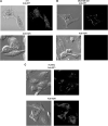
Anti-NP1 and anti-KDR peptide binding. Tumour and endothelial cells were incubated with 5-(6)-carboxyfluorescein-labelled anti-NP1 and anti-KDR peptides on chamber slides for 18 h. Peptide binding to the VEGF receptors, NP-1 and KDR, was examined on 4T1 (A), MDA-MB-231 (B) and HUVEC (C) cells by confocal microscopy (× 400 magnification). Images are representative of a scan zoom of between 1- and –4.2-fold.
Figure 3
Figure 3
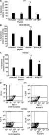
Effect of anti-NP1 and anti-KDR peptides on tumour cell and endothelial cell apoptosis. Murine 4T1 (A), human MDA-MB-231 (B) tumour cells and HUVEC endothelial cells (C) were treated with anti-NP1 and anti-KDR peptides or a ‘scrambled’ control peptide for 24 h. Annexin V binding to phosphatidylserine residues on the suface of apoptotic cells was assessed by flow cytometry and the percentage apoptotic cells was expressed relative to controls (mean±s.e.m) from three independent experiments (*P<0.05 vs untreated cells, $P<0.05 vs control peptide). Representative dot-plots demonstrating Annexin V-positive apoptotic cells are shown in the lower right-hand quadrant following treatment of murine 4T1 tumour cells (top panel) and HUVECs (lower panel) with a scrambled control peptide and anti-NP1 peptide (D).
Figure 4
Figure 4
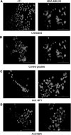
Hoechst staining of apoptotic tumour cells. 4T1 and MDA-MB-231 tumour cells were incubated for 24 h with control peptide (B), anti-NP1 (C) and anti-KDR (D) peptides. Following incubation, cells were stained with Hoechst 33342 (1 μg ml−1) in methanol and examined under UV light (× 400 magnification). Treatment of 4T1 and MDA-MB-231 tumour cells with anti-NP1 peptides induced apoptosis, as seen by bright fluorescing cells relative to control peptide and cells treated with anti-KDR peptide.

References

    1. Bachelder RE, Crago A, Chung J, Wendt MA, Shaw LM, Robinson G, Mercurio AM (2001) Vascular endothelial growth factor is an autocrine survival factor for neuropilin-expressing breast carcinoma cells. Cancer Res 61: 5736–5740 - PubMed
    1. Bachelder RE, Lipscomb EA, Lin X, Wendt MA, Chadborn NH, Eickholt BJ, Mercurio AM (2003) Competing autocrine pathways involving alternative neuropilin-1 ligands regulates chemotaxis of carcinoma cells. Cancer Res 63: 5230–5233 - PubMed
    1. Bachelder RE, Wendt MA, Mercurio AM (2002) Vascular endothelial growth factor promotes breast carcinoma invasion in an autocrine manner by regulating the chemokine receptor CXCR4. Cancer Res 62: 7203–7206 - PubMed
    1. Binetruy-Tournaire R, Demangel C, Malavaud B, Vassy R, Rouyre S, Kraemer M, Plouet J, Derbin C, Perret G, Mazie JC (2000) Identification of a peptide blocking vascular endothelial growth factor (VEGF)-mediated angiogenesis. EMBO J 7: 1525–1533 - PMC - PubMed
    1. Folkman J (1995) Angiogenesis in cancer, vascular, rheumatoid and other diseases. Nat Med 1: 27–30 - PubMed

Publication types

MeSH terms

Substances