Review
. 2005 Feb;97(2):147-72.
doi: 10.1042/BC20040058.

Virus factories: associations of cell organelles for viral replication and morphogenesis

Affiliations
Review

Virus factories: associations of cell organelles for viral replication and morphogenesis

Reyes R Novoa et al. Biol Cell. 2005 Feb.

Abstract

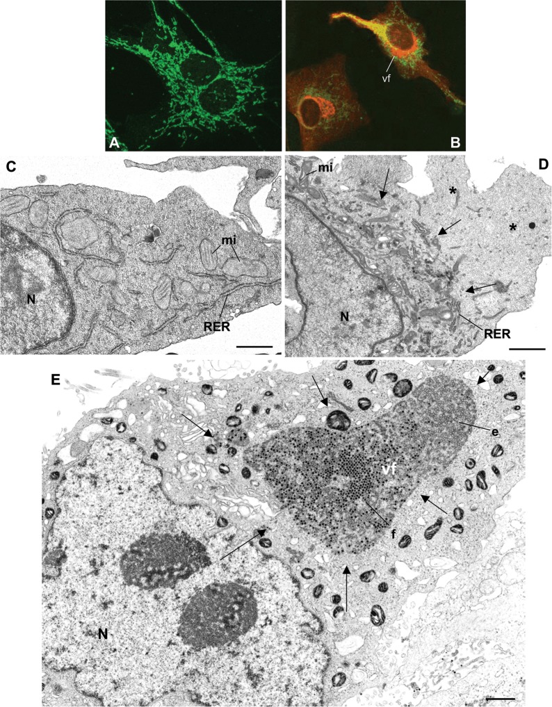
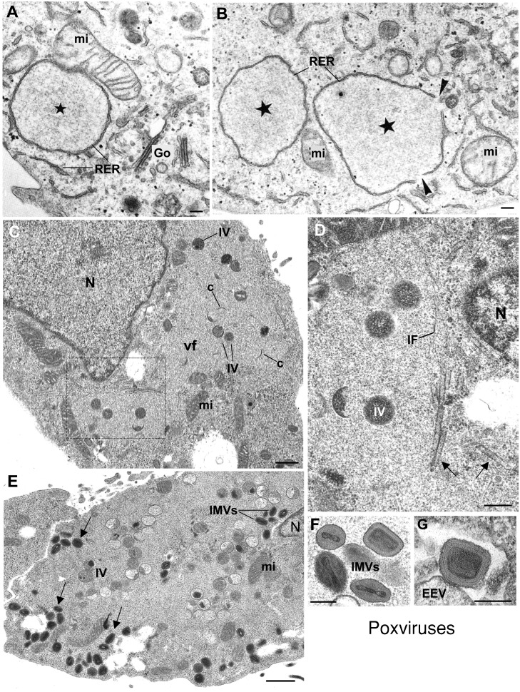
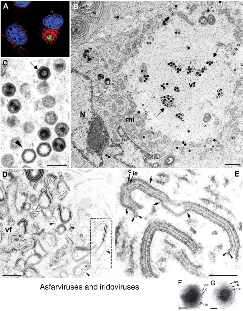
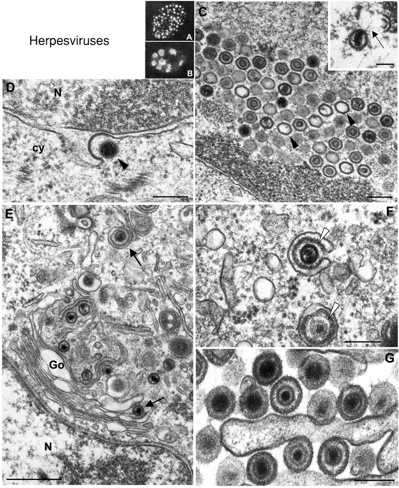
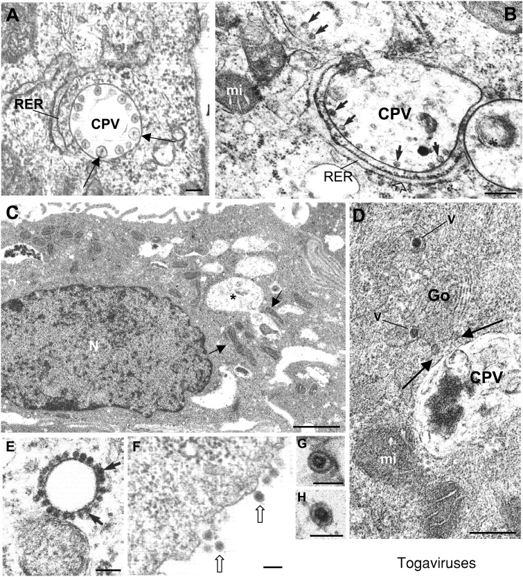
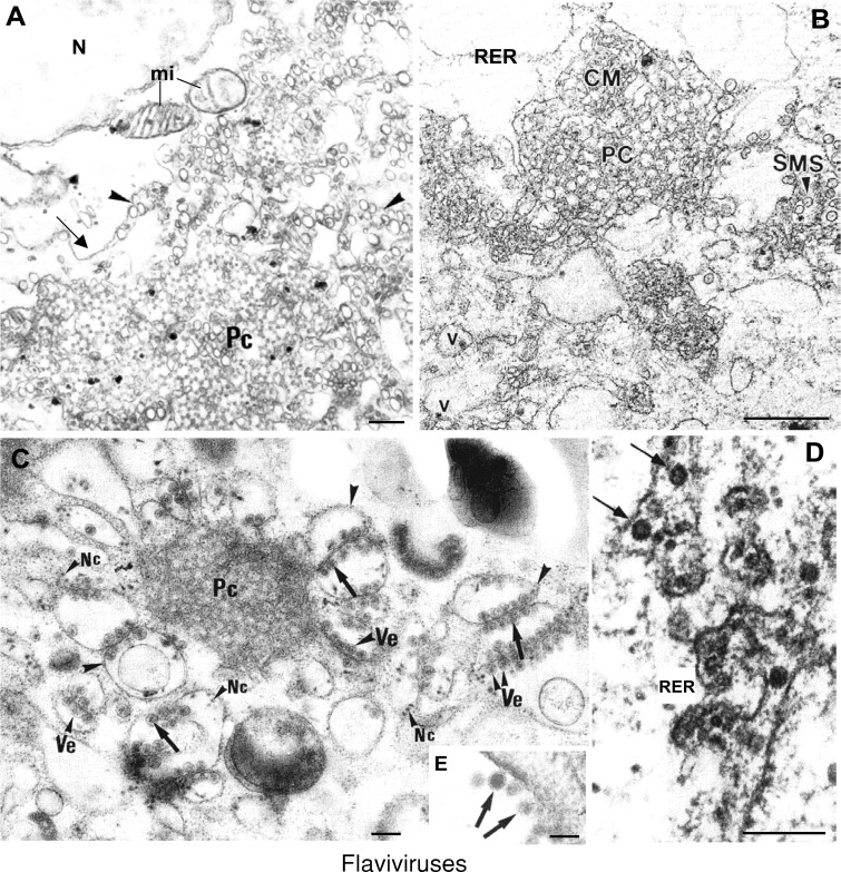
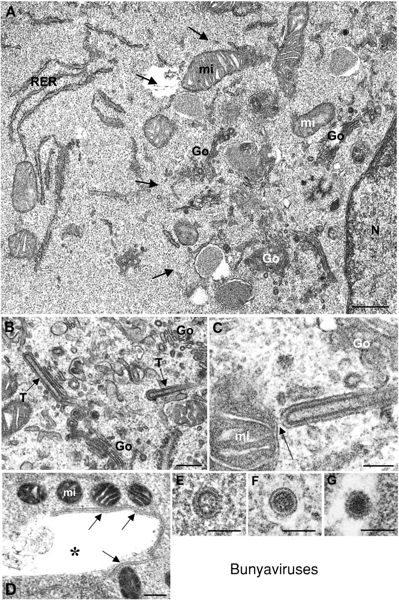
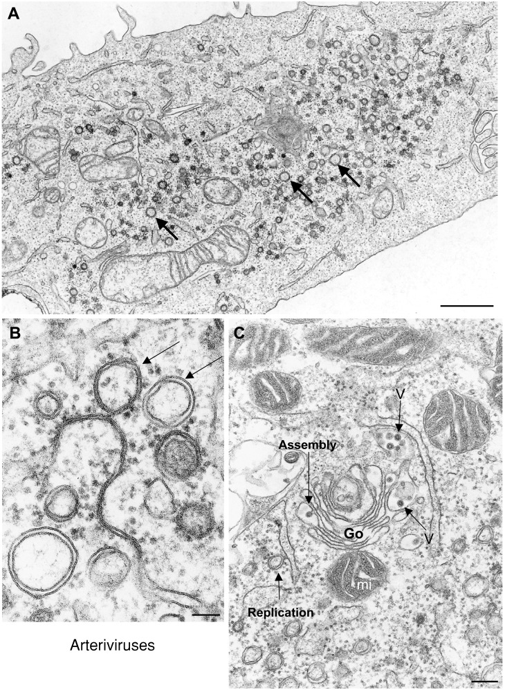
Genome replication and assembly of viruses often takes place in specific intracellular compartments where viral components concentrate, thereby increasing the efficiency of the processes. For a number of viruses the formation of 'factories' has been described, which consist of perinuclear or cytoplasmic foci that mostly exclude host proteins and organelles but recruit specific cell organelles, building a unique structure. The formation of the viral factory involves a number of complex interactions and signalling events between viral and cell factors. Mitochondria, cytoplasmic membranes and cytoskeletal components frequently participate in the formation of viral factories, supplying basic and common needs for key steps in the viral replication cycle.


Figures

Figure 1
Figure 1
Structural changes in virus‐infected cells during formation of viral factories Confocal microscopy of uninfected BHK‐21 cells (A) shows normal distribution of mitochondria (green). (B) In bunyavirus‐infected cells, mitochondria (green) have moved to the perinuclear area and surround the Golgi region (red) where the viral factory (vf) is built. Golgi complex was labelled with an anti‐giantin antibody (kindly provided by Dr. M. Renz, Institute of Immunology and Molecular Genetics, Karlsruhe, Germany) while mitochondria were labelled with Mitotracker green (Molecular Probes, Inc.). Ultrastructural analysis shows that the characteristic distribution of organelles in control cells (C) changes completely in infected cells (D) where organelles move to the perinuclear region (arrows), building a large factory and leaving the peripheral areas almost empty (asterisks). (E) In reovirus‐infected cells the vf occupies a large, but apparently more restricted area (marked with arrows) where empty (e) and full (f) viral particles are seen. N, nucleus; mi, mitochondria. Bar=0.5 μm (C) or 1 μm (D and E).
Figure 2
Figure 2
Structural changes in viral factories of VV‐infected cells (A and B) Replication complexes (stars) are rapidly formed in the perinuclear region at early times post‐infection. They are enclosed by elements of the RER. Mitochondria (mi) attach to these membranes, which start to open up (arrowheads in B) at the end of the replication phase. (C) When assembly starts, the viral factory (vf) looks like a wide area of low electron density where viral crescents (c) and immature viruses (IV) start to be distinguished. (D) Higher magnification of the area marked in (C), that has been rotated by 90°. Tubular elements of unknown identity (arrows) and IFs surround the factory. (E) At late times post‐infection the factory is filled with both IVs and IMVs, as well as mitochondria. Arrows point to mature virions that have abandoned the perinuclear region to reach the cell periphery before exit. (F and G) Higher magnification electron micrographs showing the two vaccinia infectious forms: the IMV and the EEV, respectively. V V‐infected HeLa cells were kindly provided by Dr. M. Esteban (Centro Nacional de Biotecnología, CSIC, Madrid). (A) and (B) reprinted from Molecular Biology of the Cell (Mol. Biol. Cell 2001 12: 2031–2046) with permission by the American Society for Cell Biology. N, nucleus. Bars=200 nm in A, B, D, F and G; 0.5 μm in C and E.
Figure 3
Figure 3
Factories of asfarviruses and iridoviruses Confocal microscopy shows the factories of ASFV (A) surrounded by a vimentin cage (red) that encloses viral proteins (green; nuclei are stained in blue). (B) Transmission electron microscopy shows the ultrastructure of factories for the related iridoviruses. Numerous mitochondria (mi) accumulate around the factory (vf), where viral particles start to be seen (arrows). (C) Higher magnification shows viral particles at different stages, such as empty capsids (arrowhead) and full viral particles (arrow), the latter containing the viral DNA. (D and E) show modified membranes that accumulate in viral factories (vf) induced by ASFV. These membranes come from the RER and originate the icosahedral viral core (c) and the inner envelope (ie). Extracellular virions (F and G) have an additional outer envelope (oe) taken from the plasma membrane. (A) Reproduced from The Journal of Cell Biology, 2001, 153, 449–455 with copyright permission of the Rockefeller University Press. (DG) Reproduced from Andrés et al. (1998) J. Virol. 72, 8988–9001 with copyright permission of the American Society for Microbiology. N, nucleus. Bars=1 μm in B; 250 nm in C; 0.5 μm in D; 100 nm in E; 50 nm in F and G.
Figure 4
Figure 4
Nuclear and cytoplasmic factories of herpesviruses (A) and (B) show the intranuclear location of herpesvirus prereplicative and replicative compartments, respectively, as visualized by immunofluorescence of the viral protein ICP8. (A) Vero cell infected with wild‐type virus in the presence of the viral polymerase inhibitor PAA. (B) Vero cell infected with the wild‐type virus in the absence of PAA. (C) At the ultrastructural level intranuclear empty capsids (arrowheads), as well as capsids packaging DNA (inset), are seen at later times post‐infection. (D) Release of the primary enveloped viral particle from inside the perinuclear space (arrowhead) into the cytoplasm (cy) by fusion of the primary envelope with the outer lamella of the nuclear membrane. (E) Secondary envelopment of nucleocapsids (arrows) in the trans‐Golgi area. (F) Tegumentation adds a layer of viral proteins (arrowheads) to the capsid that acquire an envelope from trans‐Golgi membranes. (G) Extracellular virions after release from cells. (A and B) reproduced from Liptak et al. (1996) J. Virol. 70, 1759–1767 with copyright permission of the American Society for Microbiology. Inset in (C) reproduced from Granzow et al. (1997) J. Virol. 71, 2072–2082, with copyright permission of the American Society for Microbiology. N, nucleus; Go, Golgi. Bars=200 nm in C, D, F, and G; 100 nm in the inset of C; 500 nm in E.
Figure 5
Figure 5
Factories of togaviruses Replication complexes of alphaviruses (A) and RUBV (B) are anchored (arrows) in the internal membrane of modified lysosomes and endosomes known as CPVs surrounded by RER cisternae. (C) Mitochondria (arrows) are seen around CPVs (asterisks) in RUBV‐infected cells. (D) Golgi stacks (Go) containing viruses (V) are seen in contact (arrows) with CPVs of RUBV‐infected cells. (E) Capsids of alphaviruses (arrows) are assembled in the cytoplasm and transported to the plasma membrane before envelopment and release to the extracellular medium (F) where extracellular virions have an homogeneously dense interior (arrows). (G and H) show extracellular mature RUBV particles that exhibit a central core separated from the envelope. (A and F) reproduced from Zhao et al. (1994) EMBO J. 13, 4204–4211, with copyright permission of EMBO J.; (B) Reprinted from Virology, 240, Magliano, Marshall, Bowden, Vardaxis, Meanger and Lee, Rubella virus replication complexes are virus modified lysosomes, pp. 57–63, Copyright (1998), with permission from Elsevier. (C, D, G and H) Reprinted from Virology, 312, Risco, Carrascosa and Frey, Structural maturation of rubella virus in the Golgi complex, pp. 261–269, Copyright (2003), with permission from Elsevier. (E) Reprinted from Virology, 265, Lee, Marshall and Bowden, Localization of rubella virus core particles in Vero cells, pp. 110–119, Copyright (1999), with permission from Elsevier. N, nucleus; mi, mitochondria. Bars=100 nm in A, B, E and F; 1 μm in C; 200 nm in D; 60 nm in G and H.
Figure 6
Figure 6
Factories of flaviviruses Rearrangement of membranes in the perinuclear region of a Vero cell infected by Kunjin virus. (A) Ordered membranes (referred to as paracrystalline arrays or Pc) and numerous vesicles (arrowheads) are surrounded by distended ER (arrows) and frequently associated with mitochondria (mi). (B) Kunjin virus‐induced Pcs in infected Vero cells are found associated with smooth convoluted membranes (CM) and smooth membrane vesicle‐like structures (SMS). Potential mature virus particles (v) are observed close to these virus‐induced membranous structures. (C) West Nile (Sarafend) virus‐induced paracrystals (Pc) and vesicles (Ve) (sample processed by cryofixation and cryosubstitution). Vesicles containing dense cores are observed around Pc membranes. Membrane bags (arrowheads) have potential nucleocapsids (Nc) associated. (D) Recombinant subviral particles of Tick‐borne encephalitis virus (formed by budding of envelope proteins in the absence of core) observed in the lumen of the RER (arrows) before their transport along the secretory pathway. (E) Extracellular mature particles of West Nile (Sarafend) virus released by infected Vero cells. (A) Reprinted from Hong and Ng (1987) Arch. Virol. 97, 115–121, with permission from Springer—Verlag. (B) Reproduced from Westaway et al. (1997) J. Virol. 71, 6650–6661, with copyright permission of the American Society for Microbiology. (C and E) Reproduced from J. Virol. Methods, 49, Ng, Teong and Tan, Cryosubstitution technique reveals new morphology of flavivirus‐induced structures, pp. 305–314, Copyright (1994), with permission from Elsevier. (D) Reproduced from Lorenz et al. (2003) J. Virol. 77, 4370–4382, with copyright permission of the American Society for Microbiology (original image by Jürgen Kartenbeck). Bars=200 nm in A, C, D; 0.5 μm in B; 50 nm in E.
Figure 7
Figure 7
Factories of bunyaviruses Factories are assembled in the perinuclear area (A), where Golgi stacks (Go) and mitochondria (mi) are seen together. RER cisternae, that are mainly excluded from the factory, are seen in peripheral areas. (B and C) Low and high‐magnification views of viral tubes (T) built in Golgi membranes and frequently seen in contact with mitochondria (arrow in C). (D) Direct contacts between mitochondria and Golgi membranes are also seen (arrows), even after Golgi disruption by monensin treatment. (EG) Viral forms assembled in infected cells. The immature precursor (shown in E) transforms into the second viral form (F) in a trans‐Golgi dependent manner. Infectious extracellular virion is shown in (G). It acquires the final structure during exit from the cell. (C and EF) reproduced from Salanueva et al. (2003) J. Virol. 77, 1368–1381, with copyright permission of the American Society for Microbiology. N, nucleus. Bars=0.5 μm in A; 200 nm in B and D; 100 nm in C, and EG.
Figure 8
Figure 8
Factories of arteriviruses Cells infected with arteriviruses (A and B) contain large amounts of double‐membranes vesicles (arrows) that represent the replication complexes of the virus and derive from the ER. (C) Replication sites are close to assembly sites in Golgi stacks (Go), where viral particles are distinguished (V). These areas are also surrounded by mitochondria (mi). Bars=1 μm in (A); 100 nm in (B); 200 nm in (C).

Similar articles

Cited by

References

    1. Andrés G. García‐Escudero R. Simón‐Mateo C. Viñuela E. African swine fever virus is enveloped by a two‐membraned collapsed cisterna from the endoplasmic reticulum J. Virol. 1998. 72 8988–9001. - PMC - PubMed
    1. Andrés G. García‐Escudero R. Salas M.L. Rodríguez J.M. Repression of African swine fever virus polyprotein pp220‐encoding gene leads to the assembly of icosahedral core‐less particles J. Virol. 2002. 76 2654–2666. - PMC - PubMed
    1. Baxter J. Merkenschlager M. Fisher A.G. Nuclear organisation and gene expression Curr. Opin. Cell Biol. 2002. 14 372–376. - PubMed
    1. Beatch M.D. Hobman T.C. Rubella virus capsid associates with host cell protein p32 and localizes to mitochondria J. Virol. 2000. 74 5569–5576. - PMC - PubMed
    1. Bienz K. Egger D. Troxler M. Pasamontes L. Structural organization of poliovirus RNA replication is mediated by viral proteins of the P2 genomic region J. Virol. 1990. 64 1156–1163. - PMC - PubMed

Publication types

LinkOut - more resources