. 2005 Jun 1;388(Pt 2):407-18.
doi: 10.1042/BJ20041556.

The CYP2B2 phenobarbital response unit contains binding sites for hepatocyte nuclear factor 4, PBX-PREP1, the thyroid hormone receptor beta and the liver X receptor

Affiliations

The CYP2B2 phenobarbital response unit contains binding sites for hepatocyte nuclear factor 4, PBX-PREP1, the thyroid hormone receptor beta and the liver X receptor

Marie-Josée Beaudet et al. Biochem J. .

Abstract

A 163 bp enhancer in the CYP2B2 5' flank confers PB (phenobarbital) inducibility and constitutes a PBRU (PB response unit). The PBRU contains several transcription factor binding sites, including NR1, NR2 and NR3, which are direct repeats separated by 4 bp of the nuclear receptor consensus half-site AGGTCA, as well as an ER (everted repeat) separated by 7 bp (ER-7). Constitutive androstane receptor (CAR)-RXR (retinoic X receptor) heterodimers are known to bind to NR1, NR2 and NR3. Electrophoretic mobility-shift analysis using nuclear extracts from livers of untreated or PB-treated rats revealed binding of several other proteins to different PBRU elements. Using supershift analysis and in vitro coupled transcription and translation, the proteins present in four retarded complexes were identified as TRbeta (thyroid hormone receptor beta), LXR (liver X receptor), HNF-4 (hepatocyte nuclear factor 4) and heterodimers of PBX-PREP1 (pre-B cell homoeobox-Pbx regulatory protein 1). LXR-RXR heterodimers bound to NR3 and TRbeta bound to NR3, NR1 and ER-7, whereas the PBX-PREP1 site is contained within NR2. The HNF-4 site overlaps with NR1. A mutation described previously, GRE1m1, which decreases PB responsiveness, increased the affinity of this site for HNF-4. The PBRU also contains a site for nuclear factor 1. The PBRU thus contains a plethora of transcription factor binding sites. The profiles of transcription factor binding to NR1 and NR3 were quite similar, although strikingly different from, and more complex than, that of NR2. This parallels the functional differences in conferring PB responsiveness between NR1 and NR3 on the one hand, and NR2 on the other.


Figures
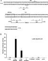
Figure 1
Figure 1. DNA sequence of rat CYP2B2 and activation by CAR of PBRU- or PBREM-driven reporter gene transcription
(A) DNA sequence of the rat CYP2B2 PBRU showing the positions of putative transcription factor binding sites. The vertical lines at nt positions −2235 and −2185 delimit the 51 bp PBREM sequence. The underlined bases in the GRE-like site are those mutated in the GRE1m1 mutation (ACA→GTG) [10,14]. (B) Activation by CAR of PBRU- or PBREM-driven reporter gene transcription. Human hepatoma HepG2 cells were cultured, and the experiments were conducted as described in the Experimental section. The corrected luciferase activity obtained for the (−120) construct co-transfected with pCMX was set at 1. The results shown are average values for three independent experiments. The error bars show the S.D. pGL3, the pBL3 basic vector; X-PBRU, a luc reporter derived from pGL3 carrying 2.5 kb of the CYP2B2 5′ flank including the PBRU in the natural sequence context; X-PBREM, identical with the X-PBRU, except that the PBRU was replaced by the PBREM; X del-PBRU, identical with X-PBRU, except that the PBRU was deleted; (−120) PBRU, the pGL3 basic vector carrying the PBRU directly upstream of the 120 bp CYP2B2 basic promoter; (−120) PBREM identical with (−120) PBRU, except that the PBRU was replaced by the PBREM; (−120), the pGL3 basic vector carrying the 120 bp CYP2B2 promoter.
Figure 2
Figure 2. HNF-4 binds to the putative GRE sequence adjacent to NR1 and the GRE1m1 mutant sequence has a higher affinity for the HNF-4 binding site
The EMSA analysis using nuclear extract from livers of untreated (C) or PB-treated (PB) rats was conducted as described in the Experimental section. PB-treated rats were injected intraperitoneally with PB daily for 3 days at a dose of 75 mg/kg. (A) EMSA and competition analyses. Unlabelled competitor oligos, when present, were at 100 ng per assay. (B) Supershift assays were conducted as described in the Experimental section. sc, supershifted complex. (C) HNF-4 obtained by coupled in vitro transcription and translation of pBluescript-HNF-4α, conducted as described in the Experimental section, binds to the HNF-4 oligo and to the GRE1m1 oligo. sc, supershifted complex; ns, complex due to proteins present in the reticulocyte extract. (D) Comparison of the sequences of the wild-type HNF-4 site that overlaps the NR1A half-site with that of the GRE1m1 mutant sequence (the mutated bases are underlined) and with the consensus of Rajas et al. [33]. (E) DNA sequence of the rat CYP2B2 PBRU showing the positions of transcription factor binding sites identified in the present study. The HNF-4 site shares a half-site with NR1 as shown. The PBX–PREP1 binding site includes the NR2 spacer and the NR2B half-site, as shown. TRβ binds to the NR1 and NR3 DR-4 sites and to the ER-7 site as shown, but not to the NR2 DR-4 site. LXR binds to the NR3 DR-4 site as shown, but not detectably to NR1 or NR2.
Figure 3
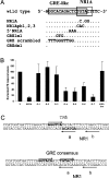
Figure 3. Assays of PB responsiveness in primary hepatocytes conferred by various mutants affected in the region of the overlapping HNF-4/NR1 site or in NR2
(A) Sequences of the mutants affected in the region of the overlapping HNF-4/NR1 site. Only the upper strand is shown. NR1A, the inactivating half-site mutation used previously [14]; NR1Apb1,2,3, an additional NR1A half-site mutation; 5′NR1A, a mutation changing 3 bp on the upstream side of NR1; GRE1m1, the GRE mutant characterized previously [10,14]; GRE scrambled, a mutation in which the GRE-like sequence was scrambled; GREdel, a mutation in which 10 bp of the GRE-like sequence was deleted. (B) Assays for PB responsiveness by transfection of primary rat hepatocytes with mutant plasmids in which the PBRU was in the natural sequence context. X, the wild-type sequence; NR1A, the NR1A half-site mutation [14]; G-1, 5′NR1A; G-2, GRE scrambled; G-3, GREdel; G-4, the GRE-like sequence was converted into a GRE consensus sequence [see (C)]; G-5, GRE1m1; G-6, the GRE-like sequence was converted into a TRβ consensus sequence [see (C)]; G-7, a double mutant with GREdel and an NR1B half-site mutation [14]; NR2-Sm, a spacer mutation of NR2 (see Figure 4). The results were normalized by comparison with the fold-induction of the X construct in parallel assays with the same hepatocyte preparation. Results were derived from duplicate assays using hepatocytes of four to nine rats per construct. The average values for the fold induction by PB varied from 27 to 54 in these experiments, a similar range to that observed previously with this system [14]. The error bars show the S.D. By Student's unpaired two-tailed one sample t test assuming unequal variance, the X construct does not differ significantly from G-1, G-3 or NR2-Sm (P≥0.4) or from G-2 (P=0.051), but differs from all the others (P<0.001). By Student's unpaired two-tailed two sample t test assuming unequal variance, G-1, G-2 and G-3 are not significantly different from each other at the 95% confidence level; nor are G-4, G-5 and G-6 or NR1A and G-7; NR1A and G-7 are different from G-4 to G-6 (P<0.01) and from G-1 to G-3 (P<0.001); and G-4 to G-6 are different from G-1 to G-3 (P<0.01). (C) The sequences, in bold characters, of the GRE consensus and the TRβ consensus mutants. Both mutant sequences overlap the NR1A half-site as shown. The GRE consensus mutant sequence differs from the GRE consensus of Evans [60] at a single position. The TRβ consensus sequence differs from the perfect TREpal sequence [61] at two positions.
Figure 4
Figure 4. PBX–PREP1 heterodimers bind to a recognition site within NR2
(A) The wild-type sequence of part of the upper strand of the NR2 site showing the PBX–PREP consensus sequence (boxed), as well as the sequence of the NR2 spacer mutation (NR2Sm) and seven single-base changes introduced into NR2 oligo sequences as shown. (B) EMSA analysis using nuclear extracts from livers of untreated (C) or PB-treated (PB) rats. The long NR2 and NR2-Sm oligos had the full-length NR2 sequence plus several base-pairs on each side, and the NR2 truncated sequence was missing 1 bp of the NR2B half site (see Table 3). Competitors, when present, were at 100 ng per assay. (C) The proteins responsible for the characteristic doublet seen with NR2 were identified by supershift analysis as PBX2–PREP1 and PBX1b–PREP1. sc, supershifted complex. (D) PBX proteins and PREP1 obtained by coupled in vitro transcription and translation bound as heterodimers to the NR2 oligo. (E) Competition assays to delineate the PBX–PREP1 binding site. The sequences of the competitor oligos, present at 100 ng per assay, are provided in (A).
Figure 5
Figure 5. EMSA analysis of rat liver nuclear proteins that bind to NR3 and to NR1
(A) Supershift analysis of liver nuclear proteins from PB-treated rats that bind to NR3 using antibodies against 10 different nuclear receptors. Retarded complexes 1–4 are indicated. Supershifted complexes were visible with anti-RXR (sc1) and anti-TRβ (sc3). The anti-UR antibody disrupted the slowest moving retarded complex (complex 1), and perhaps complex 3 as well, but here it did not give a detectable supershifted complex. TR4, testicular orphan receptor 4; PPARα, peroxisome-proliferator-activated receptor α; ERα, oestrogen receptor α; COUP-TF, chicken albumin upstream-promoter transcription factor; NURR-1, Nur-related factor 1. (B) Supershift analysis of liver nuclear proteins from untreated or PB-treated rats that bind to NR3 using anti-TRβ. Complexes 1, 2, 3 and 4 are better resolved here than in (A). (C) Competition analysis of nuclear proteins from PB-treated rats binding to NR1 using 10–100 ng of unlabelled NR1, NR3 or ER-7 competitor, as shown. Retarded complexes 2a, 2b and 4 are indicated. ns, non-specific. (D) Competition analysis of liver nuclear proteins from PB-treated rats binding to NR3 using 10–100 ng of unlabelled NR1, NR3 or ER-7 competitor, as shown. Retarded complexes 1–4 are indicated. ns, non-specific. (E) Competition analysis of liver nuclear proteins from PB-treated rats binding to NR3 using 10 or 20 ng of unlabelled NR1, NR3 or ER-7 competitor as shown. Retarded complexes 1–4 are indicated. ns, non-specific. (F) Parallel EMSA analysis with NR1 and NR3 as labelled oligos using liver nuclear proteins from PB-treated rats. (G) Supershift analysis with NR3 as labelled oligo using liver nuclear proteins from PB-treated rats in the presence of anti-RXR and anti-UR, as shown. Retarded complexes 1–4 are indicated and sc1 and sc2 are the supershifted complexes generated by anti-RXR and anti-UR respectively. (H) Supershift analysis with NR1 as labelled oligo using liver nuclear proteins from PB-treated rats in the presence of anti-RXR and anti-TRβ antibodies, as shown. Retarded complex 4 is indicated and sc1 and sc3 are the supershifted complexes generated by anti-RXR and anti-TRβ respectively. (I) Competition analysis with NR1 as the labelled oligo for binding of in vitro-synthesized rat TRβ and RXR with 10–60 ng of unlabelled NR3, NR1 or ER-7 competitor, as shown. ns, retarded complex formed from proteins present in the in vitro transcription–translation reaction mixture.

Similar articles

Cited by

References

    1. Guengerich F. P. Boca Raton: CRC Press; 1987. Mammalian Cytochromes P-450, vol. 1.
    1. Nebert D. W., Russell D. W. Clinical importance of the cytochromes P450. Lancet. 2002;360:1155–1162. - PubMed
    1. Okey A. B. Enzyme induction in the cytochrome P-450 system. Pharmacol. Ther. 1990;45:241–298. - PubMed
    1. Handschin C., Meyer U. A. Induction of drug metabolism: the role of nuclear receptors. Pharmacol. Rev. 2003;55:649–673. - PubMed
    1. Handschin C., Meyer U. A. A conserved nuclear receptor consensus sequence (DR-4) mediates transcriptional activation of the chicken CYP2H1 gene by phenobarbital in a hepatoma cell line. J. Biol. Chem. 2000;275:13362–13369. - PubMed

Publication types

MeSH terms