. 2005 Jan 18:2:2.
doi: 10.1186/1742-4690-2-2.

Inhibition of early steps in the lentiviral replication cycle by cathelicidin host defense peptides

Affiliations

Inhibition of early steps in the lentiviral replication cycle by cathelicidin host defense peptides

Lars Steinstraesser et al. Retrovirology. .

Abstract

Background: The antibacterial activity of host defense peptides (HDP) is largely mediated by permeabilization of bacterial membranes. The lipid membrane of enveloped viruses might also be a target of antimicrobial peptides. Therefore, we screened a panel of naturally occurring HDPs representing different classes for inhibition of early, Env-independent steps in the HIV replication cycle. A lentiviral vector-based screening assay was used to determine the inhibitory effect of HDPs on early steps in the replication cycle and on cell metabolism.

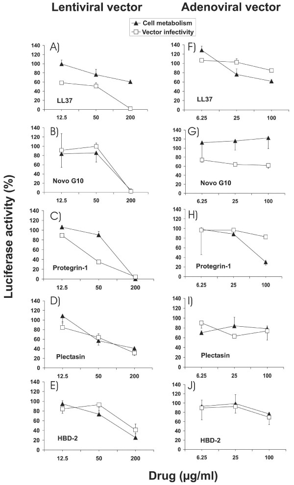
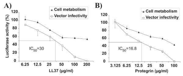
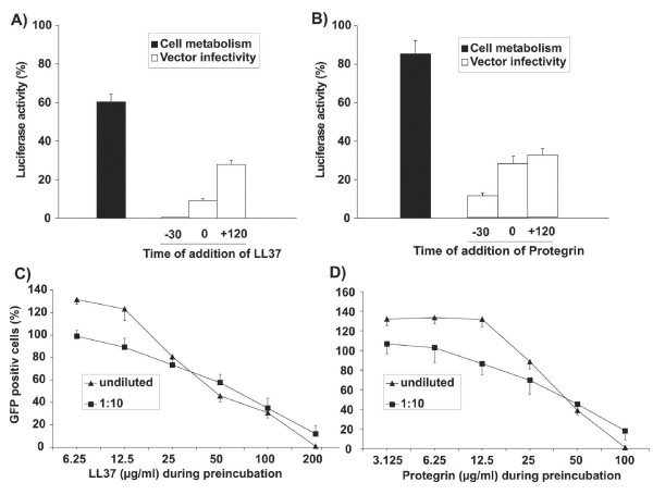
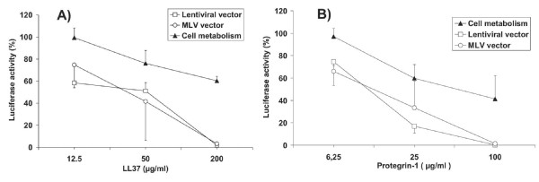
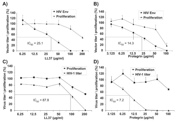
Results: Human LL37 and porcine Protegrin-1 specifically reduced lentiviral vector infectivity, whereas the reduction of luciferase activities observed at high concentrations of the other HDPs is primarily due to modulation of cellular activity and/ or cytotoxicity rather than antiviral activity. A retroviral vector was inhibited by LL37 and Protegrin-1 to similar extent, while no specific inhibition of adenoviral vector mediated gene transfer was observed. Specific inhibitory effects of Protegrin-1 were confirmed for wild type HIV-1.

Conclusion: Although Protegrin-1 apparently inhibits an early step in the HIV-replication cycle, cytotoxic effects might limit its use as an antiviral agent unless the specificity for the virus can be improved.


Figures

Figure 1
Figure 1
Comparison of different cytotoxicity assays. 293A target cells stably transduced with the luciferase gene were incubated for 48 hours in the indicated concentrations of LL37 or Protegrin-1. Viability, cell proliferation and cell metabolism of parallel cultures were assessed by a standard MTT assay, Brd-U incorporation and the luciferase assay, respectively. Values are expressed as percentage of the values obtained from cultures without HDPs. The mean and the standard deviation of triplicates are given.
Figure 2
Figure 2
Inhibitory activity of HDPs against lentiviral and adenoviral vectors. Percent luciferase activity of 293A target cells transduced in the presence of the indicated amounts of HDP with the VLΔBH lentiviral vector (A to E) or an adenoviral vector (F to J) both transferring the luciferase gene is shown. Modulation of cell metabolism was investigated in parallel by incubating 293A target cells stably transduced with a luciferase gene with the indicated amounts of HDP. The luciferase activity is expressed as percentage of the luciferase activity of cells cultured in the absence of HDP. The mean and the standard deviation of triplicates are given.
Figure 3
Figure 3
50% inhibitory concentrations of LL37 and Protegrin-1. Two-fold serial dilutions were used to determine the IC50s of LL37 (A) and Protegrin-1 for the VLΔBH vector (B). Modulation of cell metabolism was investigated in parallel by incubating 293A target cells stably transduced with a luciferase gene with the indicated amounts of HDP. The luciferase activity is expressed as percentage of the luciferase activity of cells cultured in the absence of HDP. The mean and the standard deviation of triplicates are given.
Figure 4
Figure 4
Time and concentration dependent inhibition of lentiviral vectors by LL37 and Protegrin-1. The lentiviral vector VLΔBH transferring the luciferase gene was either preincubated with 200 μg/ml of LL37 (A) or 50 μg/ml of Protegrin-1 (B) for 30 minutes (-30) or added simultaneously (0) with LL37 and Protegrin-1 to 293A target cells. Target cells were also preincubated for 120 minutes with the lentiviral vector prior to addition of LL37 and Protegrin-1. Two days after infection luciferase activities were determined as percentage of luciferase activities of cells cultured in the absence of HDPs. Cells stably transduced with the luciferase gene were also cultured in the presence and absence of LL37 and PG-1 to determine the effect of HDPs on the cell metabolism. The mean and the standard deviation of triplicates is given. A lentiviral vector transferring the GFP gene (HIV-CSCG) was incubated for 30 minutes at the indicated concentrations of LL37 (C) or Protegrin-1 (D). The vector was then added directly to 293A target cells (undiluted) or after a 1:10 dilution in medium lacking the HDPs. The number of GFP positive cells at each HDP concentration is given as percentage of GFP-positive cells of cultures transduced with diluted and undiluted vectors in the absence of HDPs. The mean and standard deviation of triplicates are shown.
Figure 5
Figure 5
Comparative analysis of inhibition of lentiviral and retroviral vector infectivity. Percent luciferase activity of 293A target cells transduced in the presence of the indicated amounts of HDP with a lentiviral vector (VLΔBH) or a retroviral vector (pRV-172) both transferring the luciferase gene is shown. Modulation of cell metabolism was investigated in parallel by incubating 293A target cells stably transduced with a luciferase gene with the indicated amounts of HDP. The luciferase activity is expressed as percentage of the luciferase activity of cells cultured in the absence of HDP. The mean and the standard deviation of triplicates of two independent experiments are shown.
Figure 6
Figure 6
Inhibitory effects of Protegrin-1 and LL37 on HIV-1 Env mediated vector entry (A, B) and HIV-1 infection (C,D). A lentiviral vector transferring the GFP gene (VGΔBH-SIN) was incubated at increasing concentrations of LL37 (A) or Protegrin-1 (B) prior to transduction of P4CCR5 cells. The vector titer is given as percentage of the titer of the vector incubated in the absence of HDPs. Wild type HIV-1 was incubated with increasing concentrations of LL37 (C) and Protegrin-1 (D). The virus titer was subsequently determined on P4CCR5 indicator cells and is expressed as percentage of the titer of the untreated HIV-1 virus stock. The toxicity of LL37 and Protegrin-1 was determined in parallel using the BrdU incorporation assay.

References

    1. Allgower M, Schoenenberger GA, Sparkes BG. Burning the largest immune organ. Burns. 1995;21:S7–47. - PubMed
    1. Steinstraesser L, Oezdogan Y, Wang SC, Steinau HU. Host defense peptides in burns. Burns. 2004;30:619–627. doi: 10.1016/j.burns.2004.05.013. - DOI - PubMed
    1. Schroder JM, Harder J. Human beta-defensin-2. Int J Biochem Cell Biol. 1999;31:645–651. doi: 10.1016/S1357-2725(99)00013-8. - DOI - PubMed
    1. Lehrer RI, Ganz T. Endogenous vertebrate antibiotics. Defensins, protegrins, and other cysteine-rich antimicrobial peptides. Ann N Y Acad Sci. 1996;797:228–239. - PubMed
    1. Boman HG. Gene-encoded peptide antibiotics and the concept of innate immunity: an update review. Scan J Immunol. 1998;48:15–25. doi: 10.1046/j.1365-3083.1998.00343.x. - DOI - PubMed

Publication types

MeSH terms

LinkOut - more resources