. 2005 Jan 19:5:8.
doi: 10.1186/1471-2407-5-8.

Upregulated expression of human neutrophil peptides 1, 2 and 3 (HNP 1-3) in colon cancer serum and tumours: a biomarker study

Affiliations

Upregulated expression of human neutrophil peptides 1, 2 and 3 (HNP 1-3) in colon cancer serum and tumours: a biomarker study

Jakob Albrethsen et al. BMC Cancer. .

Abstract

Background: Molecular markers for localized colon tumours and for prognosis following therapy are needed. Proteomics research is currently producing numerous biomarker studies with clinical potential. We investigate the protein composition of plasma and of tumour extracts with the aim of identifying biomarkers for colon cancer.

Methods: By Surface Enhanced Laser Desorption/Ionisation--Time Of Flight/Mass spectrometry (SELDI-TOF/MS) we compare the protein profiles of colon cancer serum with serum from healthy individuals and the protein profiles of colon tumours with normal colon tissue. By size exclusion chromatography, we investigate the binding of HNP 1-3 to high mass plasma proteins. By microflow we investigate the effect of HNP 1-3 on mammalian cells.

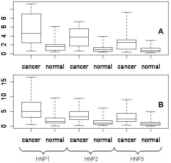
Results: Human Neutrophil Peptides -1, -2 and -3 (HNP 1-3), also known as alfa-defensin-1, -2 and -3, are present in elevated concentrations in serum from colon cancer patients and in protein extracts from colon tumours. A fraction of HNP 1-3 in serum is bound to unidentified high mass plasma proteins. HNP 1-3 purified from colon tumours are lethal to mammalian cells.

Conclusions: HNP 1-3 may serve as blood markers for colon cancer in combination with other diagnostic tools. We propose that HNP 1-3 are carried into the bloodstream by attaching to high mass plasma proteins in the tumour microenvironment. We discuss the effect of HNP 1-3 on tumour progression.


Figures

Figure 1
Figure 1
Protein profiles of serum and tissue. Representative SELDI-TOF/MS spectra of normal colon tissue (FIG. 1A) on the NP20 chip and normal serum (FIG. 1B) on the IMAC30 (Ni) chip. The two spectra differ significantly and each produce a total of 40 to 60 peaks. The majority of the peaks are present in the specified range from 2 to 10 kDa. FIG. 1C Comparison of a typical colon tumor spectrum (above) and a normal colon spectrum (below) in the range from 3 to 4 kDa. The arrows point to the three differentially expressed peptides, subsequently identified as HNP 1-3. The three peptides are expressed in normal colon tissue and the expression is upregulated in the tumor samples. The three peptides are present in normal serum, but are present in higher concentrations in colon cancer serum. The average peak intensity for the three peptides were significantly lower in serum than in tissue.
Figure 2
Figure 2
Expression of HNP 1-3 in tumors and serum. FIG.2A HNP 1-3 profiles of normal and colon tumor tissue. 40 colon tumor and 40 normal colon tissue samples were analysed on NP20 chips. Differences in mean intensities of HNP 1-3 in normal and colon tumor tissue were statistical significant at 5% level (p < 0.0005). FIG. 2B HNP profiles of normal and colon cancer serum. Serum samples (125 colon cancer and 100 normal) were analyzed on IMAC30 chips. The mean intensities were significantly different at 5% level (p < 2.2e-16). The reproducibility was significant for all three peptides in both tissue and serum. The standard deviation of HNP 1, 2 and 3 was 70%, 136% and 57% in normal tissue and 11%, 15% and 8% in tumor tissue. The standard deviation of HNP 1, 2 and 3 was 96%, 154% and 81% in normal serum and 234%, 365% and 282% in cancer serum. The boxplot show the 25th quantile, median, 75th quantile, and whiskers extent to min. and max. values.
Figure 3
Figure 3
Size exclusion study of HNP 1-3. Protein extract from tumor tissue was separated on a peptide gelfiltration column. The elution volumes of forty (unidentified) peptides was plotted against their respective mass values and an approximate elution curve was calculated. The arrows point to HNP 1-3, which were eluted in two fractions: primarily in the void volume (8 ml) together with high mass proteins (above 20 kDa) and after 14 ml together with peptides of similar mass range (2–4 kDa). We interpret this as evidence for binding between HNP 1-3 and high mass proteins.
Figure 4
Figure 4
Functional study of HNP 1-3. Normal microscopy (A&B) and flourescence microscopy (C&D) of MDCK cells. MDCK cells were exposed to calcein with (A&C) and without HNP 1-3 (B&D). By fluorescence microscopy (C&D) the cells were observed to uptake calcein only when treated with fractions containing HNP 1-3/calcein (C). Fractions containing unidentified peptides purified from colon tumors were used as negative controls together with calcein and did not stimulate the cells to uptake calcein (D). Cell islands treated with HNP 1-3 appeared diffuse and showed enlarged nuclei, indicating apoptosis (A).

References

    1. Schoen RE. The case for population-based screening for colorectal cancer. Nat Rev Cancer. 2002;2:65–70. doi: 10.1038/nrc705. - DOI - PubMed
    1. Kozak KR, Amneus MW, Pusey SM, Su F, Luong MN, Luong SA, Reddy ST, Farias-Eisner R. Identification of biomarkers for ovarian cancer using strong anion-exchange ProteinChips: potential use in diagnosis and prognosis. Proc Natl Acad Sci U S A. 2003;100:12343–12348. doi: 10.1073/pnas.2033602100. - DOI - PMC - PubMed
    1. Melle C, Ernst G, Schimmel B, Bleul A, Koscielny S, Wiesner A, Bogumil R, Moller U, Osterloh D, Halbhuber KJ, Von Eggeling F. Biomarker Discovery and Identification in Laser Microdissected Head and Neck Squamous Cell Carcinoma with ProteinChip(R) Technology, Two-dimensional Gel Electrophoresis, Tandem Mass Spectrometry, and Immunohistochemistry. Mol Cell Proteomics. 2003;2:443–452. - PubMed
    1. Petricoin EF, Ornstein DK, Paweletz CP, Ardekani A, Hackett PS, Hitt BA, Velassco A, Trucco C, Wiegand L, Wood K, Simone CB, Levine PJ, Linehan WM, Emmert-Buck MR, Steinberg SM, Kohn EC, Liotta LA. Serum proteomic patterns for detection of prostate cancer. J Natl Cancer Inst. 2002;94:1576–1578. - PubMed
    1. Ganz T. Immunology. Versatile defensins. Science. 2002;298:977–979. doi: 10.1126/science.1078708. - DOI - PubMed

Publication types

LinkOut - more resources