. 2005 Jan 25;102(4):1169-74.
doi: 10.1073/pnas.0405620102. Epub 2005 Jan 18.

EsxA and EsxB are secreted by an ESAT-6-like system that is required for the pathogenesis of Staphylococcus aureus infections

Affiliations

EsxA and EsxB are secreted by an ESAT-6-like system that is required for the pathogenesis of Staphylococcus aureus infections

Monica L Burts et al. Proc Natl Acad Sci U S A. .

Abstract

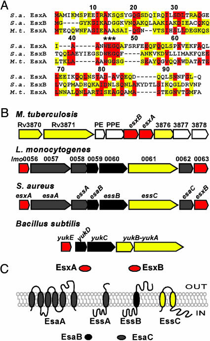
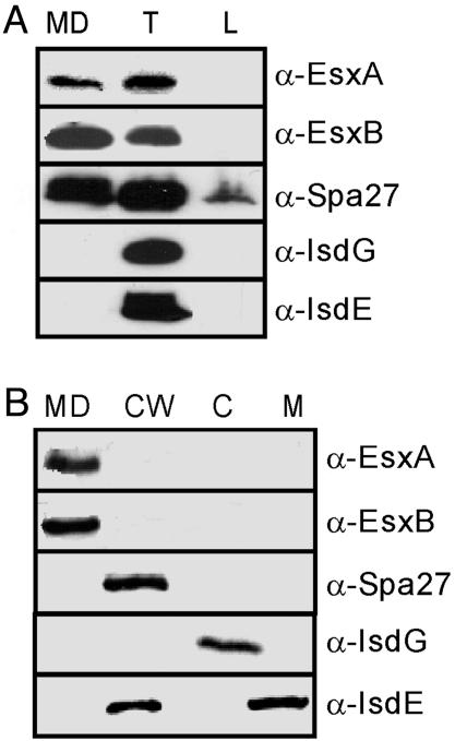
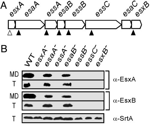
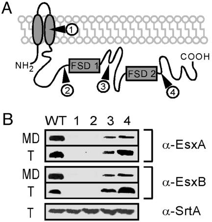
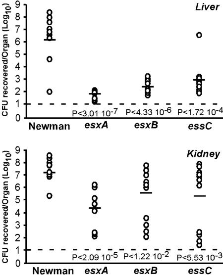
Mycobacterium tuberculosis secretes ESAT-6, a virulence factor that triggers cell-mediated immune responses and IFN-gamma production during tuberculosis. ESAT-6 is transported across the bacterial envelope by a specialized secretion system with a FSD (FtsK-SpoIIIE domain) membrane protein. Although the presence of ESAT-6-like genes has been identified in the genomes of other microbes, the possibility that they may encode general virulence functions has hitherto not been addressed. Herein we show that the human pathogen Staphylococcus aureus secretes EsxA and EsxB, ESAT-6-like proteins, across the bacterial envelope. Staphylococcal esxA and esxB are clustered with six other genes and some of these are required for synthesis or secretion of EsxA and EsxB. Mutants that failed to secrete EsxA and EsxB displayed defects in the pathogenesis of S. aureus murine abscesses, suggesting that this specialized secretion system may be a general strategy of human bacterial pathogenesis.


Figures

Fig. 1.
Fig. 1.
S. aureus ess locus encoding ESAT-6-like proteins. (A) Protein sequence alignment of S. aureus (S.a.) EsxA and EsxB with M. tuberculosis (M.t.) EsxA. M. tuberculosis and S. aureus EsxA display 20.8% identity and 25% similarity, whereas S. aureus EsxB and M. tuberculosis EsxA are 17.8% identical and 35% similar. All three proteins contain the WXG motif. (B) Comparison of the M. tuberculosis H37Rv ESAT-6 locus with the S. aureus, L. monocytogenes ess loci and the B. subtilis yuk locus. Color of genes and proteins indicates FSD factors (yellow), ESAT-6-like (red), mycobacterial genes (light gray), and staphylococcal (also in listeria or bacilli, dark gray). (C) Membrane topology or soluble character of proteins encoded by the S. aureus ess locus.
Fig. 2.
Fig. 2.
Subcellular location of EsxA and EsxB. (A) S. aureus strain Newman cultures were separated into medium (MD), total (T), and loosely wall-associated (L) fractions. Proteins were precipitated with TCA, separated on SDS/PAGE, and detected by immunoblotting with specific antibodies [α-EsxA, α-EsxB, α-Spa (protein A), α-IsdG, and α-IsdE]. (B) S. aureus strain Newman cultures were fractionated into medium (MD), cell wall (CW), cytoplasm (C), and membrane (M) compartments. TCA-precipitated proteins in each compartment were revealed by SDS/PAGE and immunoblotting.
Fig. 3.
Fig. 3.
Factors affecting the production and secretion of EsxA and EsxB. (A) Schematic drawing of the ess cluster with the position of bursa aurealis insertions (filled triangles) and esxA24 mutation (open triangles). (B) Medium (MD) and total protein fractions (T) were separated on SDS/15% PAGE and immunoblotted with α-EsxA and α-EsxB. α-SrtA is used as a control for total protein loaded.
Fig. 4.
Fig. 4.
Role of EssC in EsxA and EsxB secretion. (A) Predicted topology of EssC with insertion sites of bursa aurealis in each strain (filled triangles). EssC contains two FstK/SpoIIIE-like domains (FSD). (B) Immunoblotting of total cellular protein (T) or culture medium (MD) with α-EsxA and α-EsxB antibodies. Insertional mutations at codon 264 (mutant 1) and codon 618 (mutant 2) prevented production/secretion of EsxA and EsxB. Mutants 1–4 are strains ΦNΞ 04464 (insertion at codon 264), ΦNΞ 02191 (insertion at codon 618), ΦNΞ 02832 (insertion at codon 721), and ΦNΞ 13038 (insertion at codon 1279), respectively. α-SrtA was used as a control for the total protein loaded.
Fig. 5.
Fig. 5.
S. aureus esxA, esxB, and essC mutants are defective in the pathogenesis of murine abscesses. Ten 6-week-old BALB/c mice were injected retro-orbitally with 106 cfu for each strain. Mice were killed 4 days after infection. Kidneys and liver were removed and homogenized. Viable bacteria were counted after dilution and colony formation on tryptic soy agar. Statistical significance was examined with the Student t test, and P values were recorded. The limit of detection (solid line) was determined to be 10 cfu (101).

Similar articles

Cited by

References

    1. Rosch, J. & Caparon, M. (2004) Science 304, 1513–1515. - PubMed
    1. Campo, N., Tjalsma, H., Buist, G., Stepniak, D., Meijer, M., Veenhuis, M., Westermann, M., Muller, J. P., Bron, S., Kok, J., et al. (2004) Mol. Microbiol. 53, 1583–1599. - PubMed
    1. Chaatwal, G. S. (2002) Trends Microbiol. 10, 205–208. - PubMed
    1. Pancholi, V. & Fischetti, V. A. (1992) J. Exp. Med. 176, 415–426. - PMC - PubMed
    1. Madden, J. C., Ruiz, N. & Caparon, M. (2001) Cell 104, 143–152. - PubMed

Publication types

Substances