

Review
doi: 10.1136/hrt.2004.040337.
New insights into the pathology of inherited cardiomyopathy
Affiliations
- PMID: 15657260
- PMCID: PMC1768710
- DOI: 10.1136/hrt.2004.040337
Item in Clipboard
Review
New insights into the pathology of inherited cardiomyopathy
Heart.
2005 Feb.
No abstract available
Figures
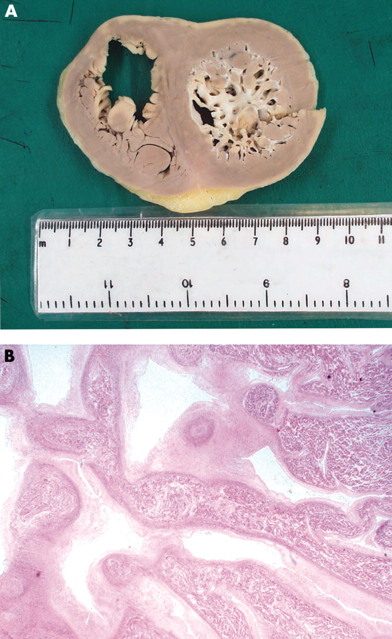
(A) Photograph of an explanted heart at mid septal level showing the gross morphological features of non-compaction in isolated LVNC. The non-compacted endocardial layer of the myocardium is comprised of excessively numerous and prominent trabeculations with deep intertrabecular recesses extending into the compacted myocardial layer imparting a distinctive “spongy” appearance. (B) Haematoxylin and eosin stained section of isolated LVNC. The non-compacted myocardial layer is comprised of prominent and elongated “finger-like” trabeculations. Within individual trabeculations there is fibrosis, which is most pronounced on the endocardial surface.
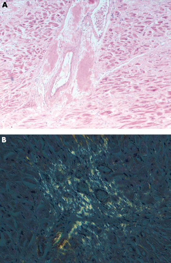
(A) Haematoxylin and eosin stained section of eosinophilic amyloid deposits within the myocardial interstitium and around blood vessels. (B) Congo red stained section of cardiac amyloidosis viewed under polarised light showing interstitial amyloid deposition with a perifibre distribution. The amyloid forms a “honeycomb” around individual myocytes and displays classical apple green birefringence.
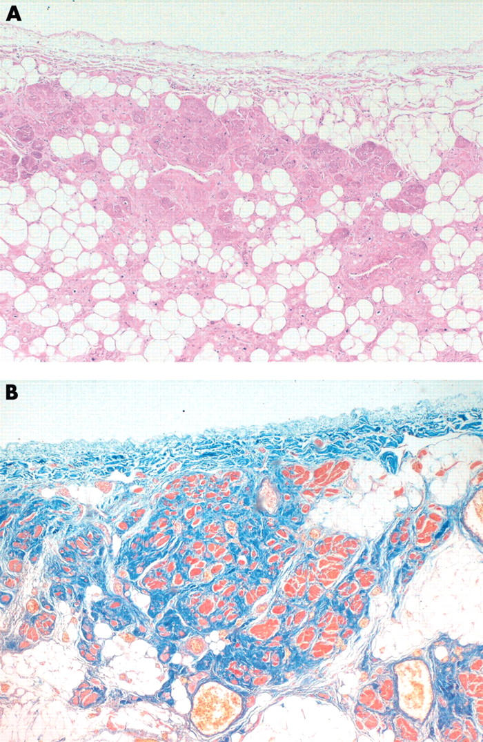
(A) Haematoxylin and eosin stained section of the “fibrofatty” or “cardiomyopathic” variant of ARVC. The fibroadipose replacement advances from the epicardium to the endocardium. There is associated myocardial atrophy and progressive myocyte loss. (B) Modified Masson’s stain demonstrating that strands and islands of residual myocytes are surrounded by fibrous tissue.
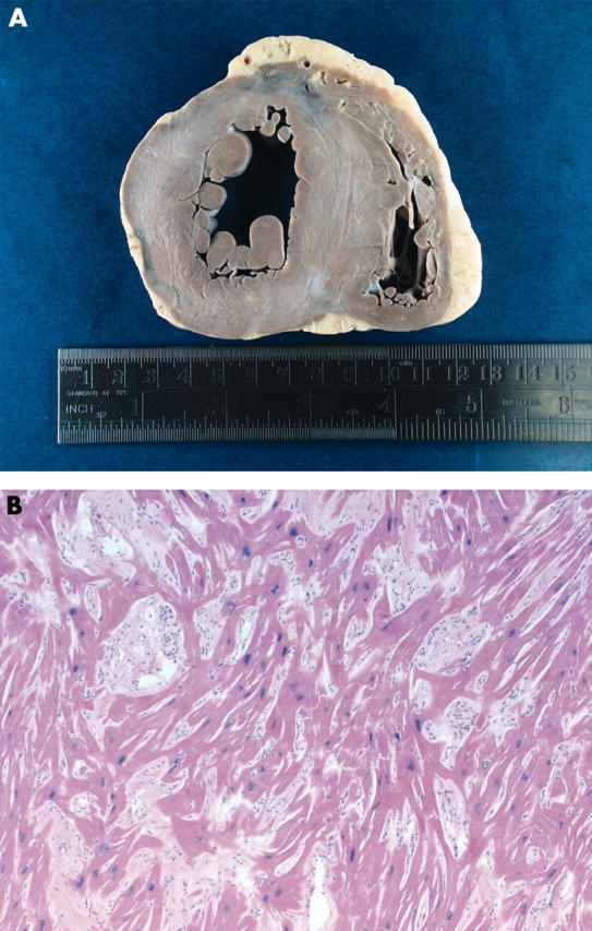
(A) Photograph of a necropsy heart at mid septal level from an individual with familial HCM. The hypertrophy involves both the free and septal walls of the left ventricle. There is disproportionate thickening of the interventricular septum, which is consistent with the asymmetrical variant of HCM. (B) Haematoxylin and eosin stained section of myocyte disarray in HCM. Disarray is characterised by hypertrophied myocytes arranged obliquely and perpendicularly around foci of interstitial collagen in a pinwheel pattern. Myocyte disarray is also accompanied by internal disorganisation of the myofibrillary architecture and nuclear changes. The latter includes hypertrophy, pleomorphism, and hyperchromasia.
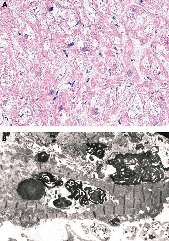
(A) Endomyocardial biopsy specimen from a patient with a clinical diagnosis of HCM. The myocytes show irregular hypertrophy and prominent sarcoplasmic vacuolisation suggestive of a myocardial metabolic storage disease. (B) Electron microscopy confirmed the presence of numerous electron dense concentric lamellar inclusions in the myocyte sarcoplasm consistent with the cardiac variant of Fabry’s disease. The diagnosis was subsequently confirmed by genotyping.
References
-
- Richardson P, McKenna WJ, Bristow M, et al. Report of the 1995 WHO/ISFC task force on the definition and classification of cardiomyopathies. Circulation 1996;93:841–42. - PubMed
-
- Towbin JA, Bowles NE. The failing heart. Nature 2002;415:227–33. ▸ An excellent up-to-date review of the identification of the genes responsible for cardiomyopathies providing further insights into the pathogenesis of these disorders. - PubMed
-
- Bowles NE, Bowles KR, Towbin JA. The “final common pathway” hypothesis and inherited cardiovascular disease. The role of cytoskeletal proteins in dilated cardiomyopathy. Herz 2000;25:168–75. - PubMed
-
- Sinagra G, Di Lenarda A, Brodsky GL, et al. Current perspective. New insights into the molecular basis of familial dilated cardiomyopathy. Ital Heart J 2001;2:280–6. ▸ A comprehensive review of the molecular genetic basis of FDCM. - PubMed
-
- Sedmera D, Pexieder T, Vuillemin M, et al. Developmental patterning of the myocardium. Anat Rec 2000;258:319–37. ▸ An excellent review article with stunning electron micrographs explaining developmental patterning of the myocardium. This article provides a unique insight into the pathology of isolated LVNC. - PubMed
Publication types
MeSH terms
LinkOut - more resources
Full Text Sources
Medical