. 2005 Jan 17;201(2):173-9.
doi: 10.1084/jem.20041212.

Prevention of UV radiation-induced immunosuppression by IL-12 is dependent on DNA repair

Affiliations

Prevention of UV radiation-induced immunosuppression by IL-12 is dependent on DNA repair

Agatha Schwarz et al. J Exp Med. .

Abstract

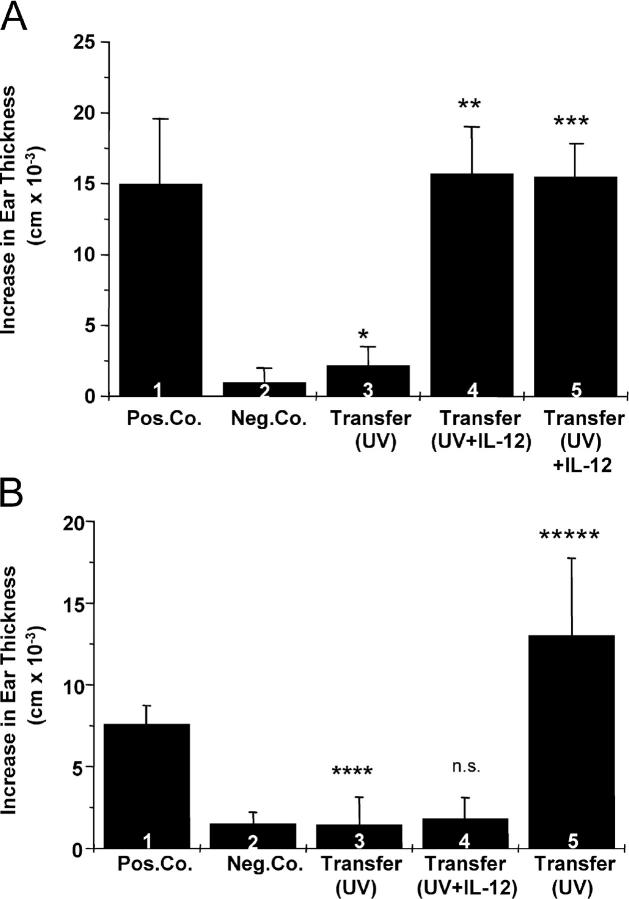
The immunostimulatory cytokine IL-12 is able to antagonize immunosuppression induced by solar/ultraviolet (UV) radiation via yet unknown mechanisms. IL-12 was recently found to induce deoxyribonucleic acid (DNA) repair. UV-induced DNA damage is an important molecular trigger for UV-mediated immunosuppression. Thus, we initiated studies into immune restoration by IL-12 to discern whether its effects are linked to DNA repair. IL-12 prevented both UV-induced suppression of the induction of contact hypersensitivity and the depletion of Langerhans cells, the primary APC of the skin, in wild-type but not in DNA repair-deficient mice. IL-12 did not prevent the development of UV-induced regulatory T cells in DNA repair-deficient mice. In contrast, IL-12 was able to break established UV-induced tolerance and inhibited the activity of regulatory T cells independent of DNA repair. These data identify a new mechanism by which IL-12 can restore immune responses and also demonstrate a link between DNA repair and the prevention of UV-induced immunosuppression by IL-12.


Figures

Figure 1.
Figure 1.
IL-12 prevents UV-induced suppression of the induction of CHS in WT but not in Xpa/ mice. C57BL/6 (A) or Xpa −/− mice (B) were treated daily with UV (1,000 and 400 J/m2, respectively) on 4 d on the back and sensitized 24 h after the last exposure through UV-exposed skin (groups 3 and 4). 5 d later, mice were challenged on the left ear and ear swelling was measured 24 h later. Group 4 received 1,000 ng of IL-12 3 h before sensitization. Positive control mice were sensitized and challenged (group 1), negative control animals were only challenged (group 2). Ear swelling is expressed as the difference (cm × 10−3, mean ± SD) between the thickness of the challenged and that of the vehicle-treated ear. *P < 0.00001 UV vs. positive control; **P < 0.005 UV vs. UV+IL-12; ***P < 0.00001 UV vs. positive control. n.s., UV vs. UV-IL-12.
Figure 2.
Figure 2.
IL-12 breaks established UV-induced tolerance in WT and Xpa/ mice. C57BL/6 (A) or Xpa −/− mice (B) were treated daily with UV (1,000 and 400 J/m2, respectively) on 4 d on the back and sensitized 24 h after the last exposure through UV-exposed skin. 14 d after the first sensitization, mice were resensitized with DNFB applied onto the abdomen (groups 3 and 4). 5 d later, mice were challenged on the right ear and ear swelling was measured 24 h later. Group 4 received 1,000 ng of IL-12 3 h before resensitization. Positive control mice were sensitized and challenged (group 1), negative control animals were only challenged (group 2). *P < 0.05 UV vs. positive control; **P < 0.05 UV vs. UV+IL-12; ***P < 0.0005 UV vs. positive control; ****P < 0.0005 UV vs. UV+IL-12.
Figure 3.
Figure 3.
IL-12 inhibits transfer of suppression by UV-induced T reg cells. Naive C57BL/6 (A) or Xpa −/− mice (B) were injected i.v. with LN cells obtained from syngeneic donors which were tolerized against DNFB by application of DNFB onto UV-exposed skin. 24 h after injection mice were sensitized against DNFB and 5 d later DNFB challenge was performed on the left ear (group 3). Mice in group 5 received 3 h before and 24 h after cell transfer 1,000 ng IL-12 i.p. Mice in group 4 received cells obtained from UV-exposed donors, which were treated with IL-12 before sensitization through UV-exposed skin, and were sensitized 24 h after cell transfer. *P < 0.001 UV vs. positive control; **P < 0.00001 UV vs. (UV+IL-12); ***P < 0.0001 UV vs. (UV)+IL-12; ****P < 0.0005 UV vs. positive control; *****P < 0.00005 UV vs. (UV)+IL-12; n.s., UV vs. (UV+IL-12).
Figure 4.
Figure 4.
IL-12 prevents UV-induced emigration of LC in WT but not in Xpa/ mice. Ears of C57BL/6 and Xpa −/− mice were exposed to 1,000 J/m2 and to 400 J/m2, respectively. 24 h later 0.5% DNFB was applied (UV+Sensi.). One group of animals received 1,000 ng IL-12 i.p. 3 h before application of DNFB (UV+Sensi.+IL-12). An additional group was only sensitized (Sensi.). Unirradiated animals served as negative controls (Control). 48 h after hapten application, ears were cut and sheet preparations performed. Sheets were stained with an anti–I-A/I-E Ab, followed by an anti–rat IgG coupled with Texas red and subjected to fluorescence microscopy.
Figure 5.
Figure 5.
IL-12 reduces the number of CPD-positive LC in LN draining UV-exposed skin in WT but not in Xpa/ mice. C57BL/6 or Xpa −/− mice were treated daily with UV on 4 d on the shaved back and sensitized 24 h after the last exposure through UV-exposed skin (UV). One group received 1,000 ng of IL-12 3 h before sensitization (UV+IL-12). As controls LN of untreated and DNFB-sensitized (Sensi.) mice were used. 48 h after sensitization CD11c-positive cells were obtained from draining LN by magnetobead separation. Cells were double stained for Langerin and CPDs and subjected to FACS analysis.

References

    1. Fisher, G.J., S.C. Datta, H.S. Halwar, Z.Q. Wang, J. Varani, S. Kang, and J.J. Voorhees. 1996. Molecular basis of sun-induced premature skin ageing and retinoid antagonism. Nature. 379:335–339. - PubMed
    1. de Gruijl, F.R., H.J. Sterenborg, P.D. Forbes, R.E. Davies, C. Cole, G. Kelfkens, H. van Weelden, H. Slaper, and J.C. van der Leun. 1993. Wavelength dependence of skin cancer induction by ultraviolet irradiation of albino hairless mice. Cancer Res. 53:53–60. - PubMed
    1. Chapman, R.S., K.D. Cooper, E.C. DeFabo, J.E. Frederick, K.N. Gelatt, S.P. Hammond, P. Hersey, H.S. Koren, R.D. Ley, F. Noonan, et al. 1995. Solar ultraviolet radiation and the risk of infectious disease. Photochem. Photobiol. 61:223–247. - PubMed
    1. Toews, G.B., P.R. Bergstresser, and J.W. Streilein. 1980. Epidermal Langerhans cell density determines whether contact hypersensitivity or unresponsiveness follows skin painting with DNFB. J. Immunol. 124:445–453. - PubMed
    1. Cooper, K.D., L. Oberhelman, T.A. Hamilton, O. Baadsgaard, M. Terhune, G. LeVee, T. Anderson, and H. Koren. 1992. UV exposure reduces immunization rates and promotes tolerance to epicutaneous antigens in humans: Relationship to dose, CD1a-DR+epidermal macrophage induction, and Langerhans cell depletion. Proc. Natl. Acad. Sci. USA. 89:8497–8501. - PMC - PubMed

Publication types

MeSH terms