

Contrasting frequencies of antitumor and anti-vaccine T cells in metastases of a melanoma patient vaccinated with a MAGE tumor antigen
- PMID: 15657294
- PMCID: PMC2212799
- DOI: 10.1084/jem.20041378
Contrasting frequencies of antitumor and anti-vaccine T cells in metastases of a melanoma patient vaccinated with a MAGE tumor antigen
Abstract
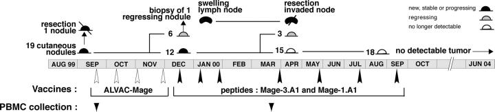
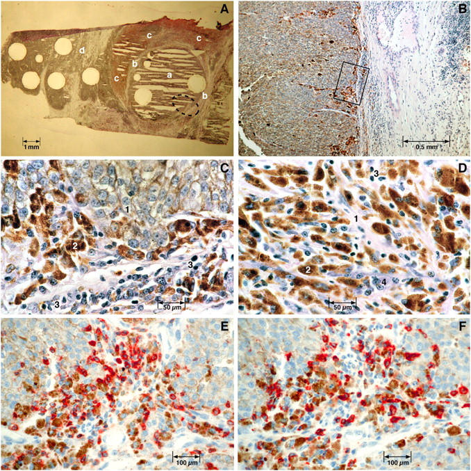
Melanoma patients have high frequencies of T cells directed against antigens of their tumor. The frequency of these antitumor T cells in the blood is usually well above that of the anti-vaccine T cells observed after vaccination with tumor antigens. In a patient vaccinated with a MAGE-3 antigen presented by HLA-A1, we measured the frequencies of anti-vaccine and antitumor T cells in several metastases to evaluate their respective potential contribution to tumor rejection. The frequency of anti-MAGE-3.A1 T cells was 1.5 x 10(-5) of CD8 T cells in an invaded lymph node, sixfold higher than in the blood. An antitumor cytotoxic T lymphocyte (CTL) recognizing a MAGE-C2 antigen showed a much higher enrichment with a frequency of approximately 10%, 1,000 times higher than its blood frequency. Several other antitumor T clonotypes had frequencies >1%. Similar findings were made on a regressing cutaneous metastasis. Thus, antitumor T cells were approximately 10,000 times more frequent than anti-vaccine T cells inside metastases, representing the majority of T cells present there. This suggests that the anti-vaccine CTLs are not the effectors that kill the bulk of the tumor cells, but that their interaction with the tumor generates conditions enabling the stimulation of large numbers of antitumor CTLs that proceed to destroy the tumor cells. Naive T cells appear to be stimulated in the course of this process as new antitumor clonotypes arise after vaccination.
Figures
References
-
- Karanikas, V., C. Lurquin, D. Colau, N. van Baren, C. De Smet, B. Lethé, T. Connerotte, V. Corbière, M.-A. Demoitié, D. Liénard, et al. 2003. Monoclonal anti-MAGE-3 CTL responses in melanoma patients displaying tumor regression after vaccination with a recombinant canarypox virus. J. Immunol. 171:4898–4904. - PubMed
-
- Lennon, G.P., J.E. Sillibourne, E. Furrie, M.J. Doherty, and R.A. Kay. 2000. Antigen triggering selectively increases TCRBV gene transcription. J. Immunol. 165:2020–2027. - PubMed
-
- Davies, H., G.R. Bignell, C. Cox, P. Stephens, S. Edkins, S. Clegg, J. Teague, H. Woffendin, M.J. Garnett, W. Bottomley, et al. 2002. Mutations of the BRAF gene in human cancer. Nature. 417:949–954. - PubMed
-
- Shimizu, J., S. Yamazaki, and S. Sakaguchi. 1999. Induction of tumor immunity by removing CD25+CD4+ T cells: a common basis between tumor immunity and autoimmunity. J. Immunol. 163:5211–5218. - PubMed
Publication types
MeSH terms
Substances
LinkOut - more resources
Full Text Sources
Other Literature Sources
Medical
Research Materials
Miscellaneous
