. 2005 Feb;125(2):171-86.
doi: 10.1085/jgp.200409076. Epub 2005 Jan 18.

Preferential phosphorylation of R-domain Serine 768 dampens activation of CFTR channels by PKA

Affiliations

Preferential phosphorylation of R-domain Serine 768 dampens activation of CFTR channels by PKA

László Csanády et al. J Gen Physiol. 2005 Feb.

Abstract

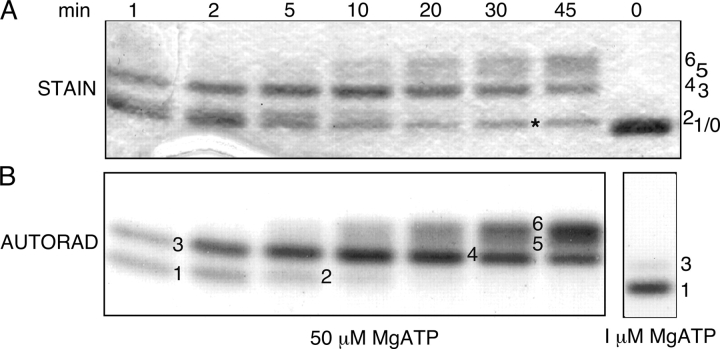
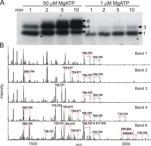
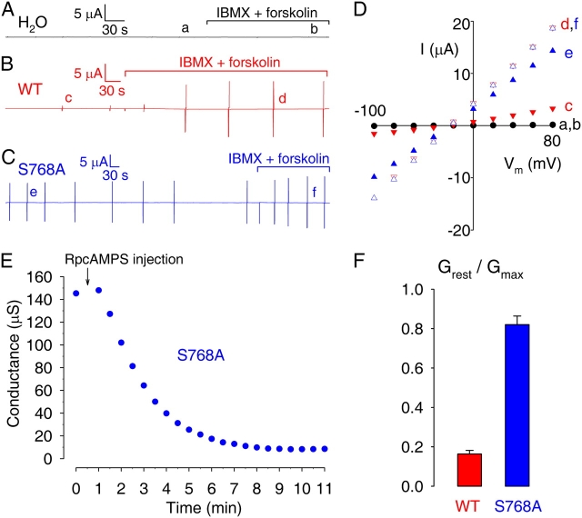
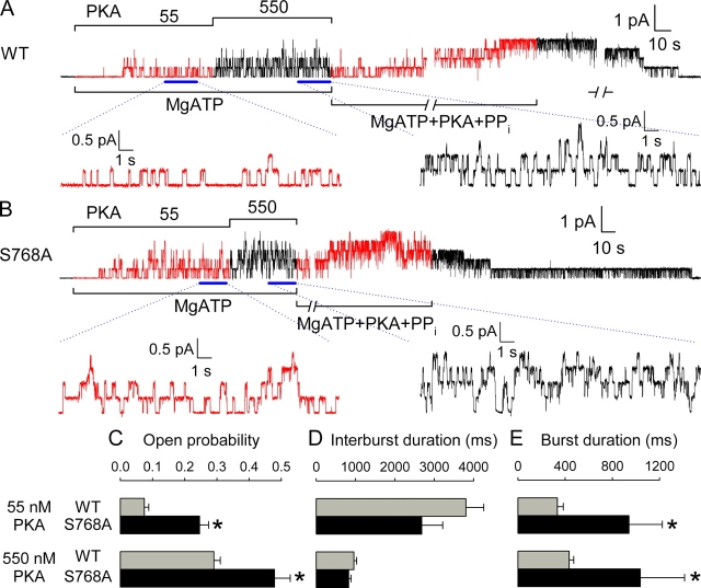
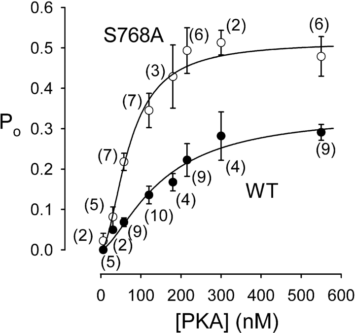
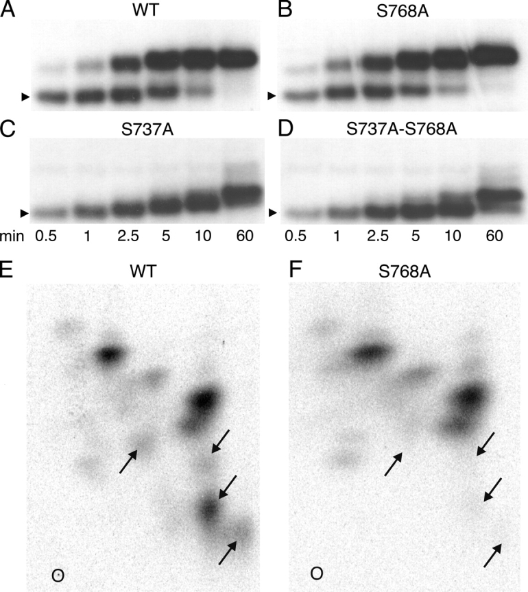
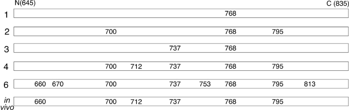
CFTR (cystic fibrosis transmembrane conductance regulator), the protein whose dysfunction causes cystic fibrosis, is a chloride ion channel whose gating is controlled by interactions of MgATP with CFTR's two cytoplasmic nucleotide binding domains, but only after several serines in CFTR's regulatory (R) domain have been phosphorylated by cAMP-dependent protein kinase (PKA). Whereas eight R-domain serines have previously been shown to be phosphorylated in purified CFTR, it is not known how individual phosphoserines regulate channel gating, although two of them, at positions 737 and 768, have been suggested to be inhibitory. Here we show, using mass spectrometric analysis, that Ser 768 is the first site phosphorylated in purified R-domain protein, and that it and five other R-domain sites are already phosphorylated in resting Xenopus oocytes expressing wild-type (WT) human epithelial CFTR. The WT channels have lower activity than S768A channels (with Ser 768 mutated to Ala) in resting oocytes, confirming the inhibitory influence of phosphoserine 768. In excised patches exposed to a range of PKA concentrations, the open probability (P(o)) of mutant S768A channels exceeded that of WT CFTR channels at all [PKA], and the half-maximally activating [PKA] for WT channels was twice that for S768A channels. As the open burst duration of S768A CFTR channels was almost double that of WT channels, at both low (55 nM) and high (550 nM) [PKA], we conclude that the principal mechanism by which phosphoserine 768 inhibits WT CFTR is by hastening the termination of open channel bursts. The right-shifted P(o)-[PKA] curve of WT channels might explain their slower activation, compared with S768A channels, at low [PKA]. The finding that phosphorylation kinetics of WT or S768A R-domain peptides were similar provides no support for an alternative explanation, that early phosphorylation of Ser 768 in WT CFTR might also impair subsequent phosphorylation of stimulatory R-domain serines. The observed reduced sensitivity to activation by [PKA] imparted by Ser 768 might serve to ensure activation of WT CFTR by strong stimuli while dampening responses to weak signals.


Figures

F<sc>igure</sc> 1.
Figure 1.
Time course of in vitro phosphorylation of R-domain peptide by PKA revealed by SDS-PAGE. Incubations were with 50 μM γ32P-MgATP for various times as indicated (0–45 min), except for the sample shown in the lower right panel, which was incubated with 1 μM γ32P-MgATP for 2 min. Samples were separated by SDS-PAGE, gels stained with Coomassie blue (A, STAIN), dried, and exposed to X-ray film (B, AUTORAD). Numbers label bands of varying mobility (stain band labeled * is an impurity). The unphosphorylated R domain (sample at 0 min) migrates as a single species of ∼28 kD (band 0, upper panel, extreme right).
F<sc>igure</sc> 2.
Figure 2.
Order of in vitro PKA phosphorylation of R-domain peptide determined by SDS-PAGE and mass spectrometry. (A) R-domain protein was incubated with 50 μM or 1 μM γ32P-MgATP. Samples were separated by SDS-PAGE and exposed to X-ray film. The gel bands indicated (numbered arrows) were excised (Band 6 was excised from the upper part of the top-most band in the 10-min lane at 50 μM MgATP, as indicated by the arrow), digested by trypsin, and the resulting proteolytic products subjected to mass spectrometric analysis. (B) MALDI-ion trap mass spectrometric analysis of the five bands indicated in A showing that serine 768 is the most readily phosphorylated residue. Pairs of peaks separated by 98 D (joined by the red and blue connecting lines) provide signatures for phosphorylated peptides (see text). The blue lines designate peptide that is singly oxidized at methionine 773 (or 721, Band 4); the red lines indicate the corresponding unoxidized peptides. The amino acid residues of these phosphopeptides are indicated above each pair of peaks. For example, 766–785 and 765–785 correspond to two alternative cleavage products of the trypsin digestion. Because the masses of phosphorylated peptides 811–830 and 766–785 are respectively 2411.110 and 2411.164 (Table I), these two species were not resolved in this experiment, but the presence of 811–830 was inferred from the change in ratio of the pair of peaks associated with this mass in Band 6 (bottom panel) compared with other bands, which contain only 766–785. The peptide 766–785 contains a single methionine, which was partially oxidized to give the two pairs of peaks seen in all five panels. As peptide 811–830 contains no methionine residue, the presence of peptide 811–830 together with peptide 766–785 is signaled by a change in the ratio of the pairs of peaks 16 D apart.
F<sc>igure</sc> 3.
Figure 3.
Membrane conductance of resting and activated oocytes expressing WT or mutant S768A CFTR. Current time courses, recorded under two-microelectrode voltage clamp, of oocytes injected with water (A), or with cRNA encoding (B) WT or (C) S768A CFTR. Membrane potential was held near the resting potential (between −40 and −20 mV). Membrane conductance was monitored at regular intervals using brief voltage steps (vertical lines); complete current/voltage (I/V) relationships were determined under resting conditions (time points a, c, e), and after activation of cellular PKA with 50 μM forskolin + 1 mM IBMX (time points b, d, f). (D) Representative I/V plots under conditions as indicated in A–C, showing steady currents measured by averaging current samples toward the ends of 75-ms steps to voltages between −100 and +80 mV. (E) Rapid reduction of resting conductance (from 145 to 8 μS) upon injection of RpcAMPS into an oocyte expressing S768A CFTR. (F) Ratios, for WT- and S768A-expressing oocytes, of membrane conductances at rest (Grest), and after maximal activation of CFTR (Gmax); conductance values were similar for oocytes injected with 2.5 or 5 ng of each cRNA, so pooled results are shown.
F<sc>igure</sc> 4.
Figure 4.
Serine 768 of WT CFTR is significantly phosphorylated in vivo in resting oocytes. MALDI-TOF (top) and MALDI-QqTOF (bottom) mass spectra of peptides obtained by trypsin digestion of WT CFTR immunoprecipitated from the membranes of resting oocytes that were lysed in the presence of phosphatase inhibitors to preserve phosphoserines present at that instant. Arrows identify phosphorylated (+P) peptides (molecular masses are listed in Table I).
F<sc>igure</sc> 5.
Figure 5.
Activation in excised patches of macroscopic WT and S768A CFTR currents by low and high concentrations of PKA catalytic subunit. (A and B) Currents recorded in patches containing hundreds of WT (A) or S768A (B) CFTR channels. No substantial current is activated in either case by 2 mM MgATP applied ∼2 min after patch excision, but 55 nM, and 550 nM, PKA catalytic subunit activate increasing amounts of current in both cases. Solid green and blue lines show single-exponential fits to the current time courses, with indicated time constants; activation/inactivation time course of endogenous Ca2+-activated Cl channel current elicited by a brief pulse of 2 mM Ca2+ was used to estimate the speed of solution exchange. (C) Fractional current activated by 55 nM PKA was significantly smaller for WT (gray bar) than S768A (black bar; *, P = 0.0024) CFTR channels; mean steady current in 55 nM PKA was divided by mean steady current in the same patch at 550 nM PKA. (D) Time constants of macroscopic current relaxations upon addition of 55 nM (top) or 550 nM (bottom) PKA, for WT (gray bars) and S768A (black bars) CFTR; activation was faster for S768A at low (*, P = 0.036), but not at high [PKA].
F<sc>igure</sc> 6.
Figure 6.
Kinetic behavior of WT and S768A CFTR channels in excised patches exposed to low and high [PKA]. Representative baseline-subtracted current traces of (A) four WT and (B) five S768A channels, recorded from excised patches in the presence of 2 mM MgATP + 55 nM or 550 nM PKA; 20-s segments (indicated by bars) under each condition are shown with 10-fold expanded time scale, below. Channels were counted by locking them in the open-burst state with 0.1 mM MgATP + 2 mM pyrophosphate (PPi) + 300 nM PKA. (C–E) Open probabilities (C), mean interburst (D) and open burst (E) durations at 55 nM (top) or 550 nM PKA (bottom), for WT (gray bars) and S768A (black bars) CFTR channels; asterisks indicate significant differences between S768A and WT (0.001 < P < 0.06).
F<sc>igure</sc> 7.
Figure 7.
Dependence on [PKA] of open probability, Po, of WT (•) and S768A (○) CFTR channels. Steady-state mean Po measured in (n) excised patches, containing few channels, during exposure to 6–550 nM PKA plus near-saturating [MgATP] (1–2 mM), is plotted against [PKA]. Lines show nonlinear least-squares fits to the Hill equation, yielding Po,max = 0.34 ± 0.06, K 0.5 = 149 ± 46 nM, nH = 1.5 ± 0.5 for WT, and Po,max = 0.51 ± 0.05, K 0.5 = 71 ± 12 nM, nH = 1.8 ± 0.5 for S768A.
F<sc>igure</sc> 8.
Figure 8.
(A–D) Time courses of in vitro phosphorylation of R-domain peptides with COOH-terminal His tags. WT (A) and mutant R-domain peptides, with Ser768 replaced by alanine (S768A; B), Ser737 replaced by alanine (S737A; C), or Ser737 and Ser768 both replaced by alanine (S737A-S768A; D), were phosphorylated with 10 nM PKA and 5 μM γ32P-MgATP for 0.5–60 min as indicated. Samples were subjected to SDS-PAGE and analyzed by autoradiography; the arrowheads indicate relative molecular mass of 28 kD. The major mobility shift (to band 3; Figs. 1 and 2) was seen in the WT and S768A peptides, but not in the S737A or S737A-S768A peptides. The kinetics of R-domain phosphorylation, as demonstrated by the mobility shifts, was little altered in the two S768A mutants. (E and F) Two-dimensional tryptic phosphopeptide maps of His-tagged WT and S768A R-domain peptides phosphorylated in vitro as in A and B, but for 0.5 min with 10 nM PKA and 50 μM γ32P-MgATP, and then subjected to SDS-PAGE and analyzed by autoradiography. The lower radioactive bands were excised, digested overnight with 50 μg/ml TPCK-trypsin, and the digests separated on thin layer cellulose plates by electrophoresis at pH 3.5 in the first dimension and ascending chromatography in the second dimension. O, 0rigin; left, positive; right, negative. Four spots (arrows) in the WT R domain map are absent from the S768A map, but no similarly striking differences are seen in the pattern or intensity of other spots.
F<sc>igure</sc> 9.
Figure 9.
Summary of R-domain serines found to be phosphorylated by mass spectrometric analysis of trypic digests of bands (1, 2, 3, 4, 6 as indicated) from SDS-PAGE gels after in vitro phosphorylation (data from Fig. 2) or of full-length WT CFTR isolated from resting oocytes (in vivo; data from Fig. 4). Ser 768 is the first site phosphorylated, and is found phosphorylated in oocytes at rest; the major mobility shift to band 3 is associated with phosphorylation of Ser 737.

Similar articles

Cited by

References

    1. Aleksandrov, A.A., X.-B. Chang, L. Aleksandrov, and J.R. Riordan. 2000. The non-hydrolytic pathway of cystic fibrosis transmembrane conductance regulator ion channel gating. J. Physiol. 528:259–265. - PMC - PubMed
    1. Aleksandrov, L., A. Mengos, X. Chang, A. Aleksandrov, and J.R. Riordan. 2001. Differential interactions of nucleotides at the two nucleotide binding domains of the cystic fibrosis transmembrane conductance regulator. J. Biol. Chem. 276:12918–12923. - PubMed
    1. Aleksandrov, L., A.A. Aleksandrov, X.B. Chang, and J.R. Riordan. 2002. The first nucleotide binding domain of cystic fibrosis transmembrane conductance regulator is a site of stable nucleotide interaction, whereas the second is a site of rapid turnover. J. Biol. Chem. 277:15419–15425. - PubMed
    1. Basso, C., P. Vergani, A.C. Nairn, and D.C. Gadsby. 2003. Prolonged nonhydrolytic interaction of nucleotide with CFTR's NH2-terminal nucleotide binding domain and its role in channel gating. J. Gen. Physiol. 122:333–348. - PMC - PubMed
    1. Borchardt, R., J. Kole, and J.A. Cohn. 1996. Phosphorylation of CFTR Ser-737 by protein kinase A. Pediatr. Pulmonol. Suppl. 13:212.

Publication types

Substances