. 2005 Jan 17;168(2):209-19.
doi: 10.1083/jcb.200408087.

Spindle-independent condensation-mediated segregation of yeast ribosomal DNA in late anaphase

Affiliations

Spindle-independent condensation-mediated segregation of yeast ribosomal DNA in late anaphase

Félix Machín et al. J Cell Biol. .

Abstract

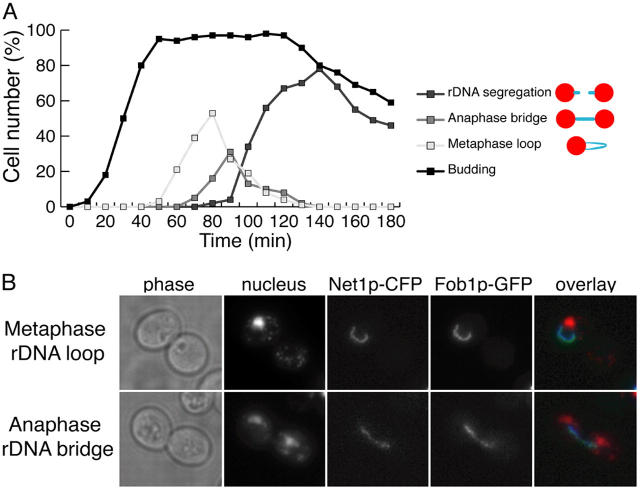
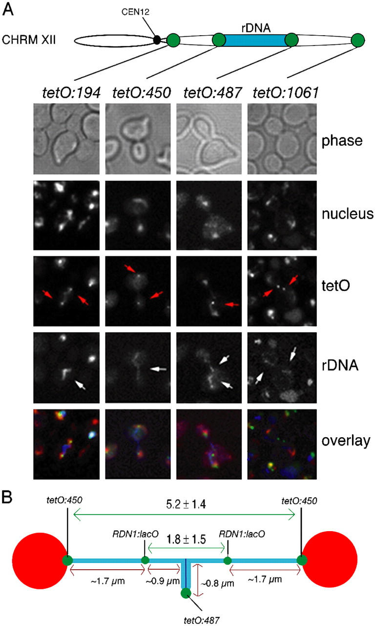
Mitotic cell division involves the equal segregation of all chromosomes during anaphase. The presence of ribosomal DNA (rDNA) repeats on the right arm of chromosome XII makes it the longest in the budding yeast genome. Previously, we identified a stage during yeast anaphase when rDNA is stretched across the mother and daughter cells. Here, we show that resolution of sister rDNAs is achieved by unzipping of the locus from its centromere-proximal to centromere-distal regions. We then demonstrate that during this stretched stage sister rDNA arrays are neither compacted nor segregated despite being largely resolved from each other. Surprisingly, we find that rDNA segregation after this period no longer requires spindles but instead involves Cdc14-dependent rDNA axial compaction. These results demonstrate that chromosome resolution is not simply a consequence of compacting chromosome arms and that overall rDNA compaction is necessary to mediate the segregation of the long arm of chromosome XII.


Figures

Figure 1.
Figure 1.
A defined “anaphase rDNA bridge” stage occurs after the rDNA loop in synchronous cultures. (A) Strain CCG1580 (Mata FOB1-GFP NET1-CFP) was arrested in G1 using α-factor and released into fresh medium at 30°C. Time-points collected every 10 min were analyzed for the organization of rDNA arrays. Categories scored are shown in the figure legend and include rDNA metaphase loops, “anaphase bridges” and segregated rDNA. (B) Strain CCG1580 (Mata FOB1-GFP NET1-CFP) was arrested in metaphase using nocodazole and released into fresh medium at 30°C. Micrographs representative of metaphase rDNA loops and anaphase rDNA bridges are shown.
Figure 2.
Figure 2.
Sister rDNA arrays unzip from centromere-proximal to centromere-distal regions during bridge the stage before segregation to opposite poles. (A) Strains CCG1326 (Mata TetR-YFP tetO:194Kb-ChrXII NET1-CFP), CCG1327 (Mata TetR-YFP tetO:450Kb-ChrXII NET1-CFP), CCG1328 (Mata TetR-YFP tetO:487Kb-ChrXII NET1-CFP), and CCG1329 (Mata TetR-YFP tetO:1061Kb-ChrXII NET1-CFP), were released from a G1 block and imaged as cells underwent segregation using nuclear separation as a reference for anaphase entry. All the cells analyzed during the bridge period showed the pattern displayed in the representative cells of each category. Chromosome tags (red arrows) and rDNA arrays (white arrows) are indicated. Sister rDNA arrays are separated at their centromeric proximal flanks yet are still connected through their telomeric flanks during the anaphase bridge period. Note that the tetO:1061 panel shows a cell already transiting from the bridge to fully segregated rDNA in order to show that sister chromosome XII right telomeres trail rDNA during the segregation. (B) Distances between tags at the rDNA bridge stage were measured for strains CCG1327 (Mata TetR-YFP tetO:450Kb-ChrXII NET1-CFP) and CCG1191 (Mata LacI-GFP RDN1:lacO TUB4-CFP NET1-CFP) after a G1 block release. The position of the lacO in the middle of the rDNA (Torres-Rosell et al., 2004) allowed to estimate the percentage of rDNA array resolved at this stage. We found it to be ∼77% (1.7 μm of resolved rDNA chromatids/3.4 μm of total rDNA chromatid length).
Figure 3.
Figure 3.
Axial compaction of rDNA arrays occurs after the anaphase bridge period and is not a consequence of nuclear envelop fission. (A) The average axial length of the rDNA arrays was measured in metaphase loops, anaphase bridges, or segregated rDNA arrays in the CCG1580 strain. Note that the average axial length of rDNA loops represents cohesed chromatids, the average length of anaphase rDNA bridges represents two chromatids that are separated at their centromeric proximal flanks but still connected through their telomeric flanks; and the average length of segregated rDNA chromatids represents that of each chromatid (i.e., the value of the anaphase rDNA bridge should be divided by a factor of two for direct comparison to the value of metaphase loops and segregated chromatids, which represent a single chromatid length). Overall, rDNA compaction takes place after the anaphase bridge period. (B) Representative micrograph of a cell in telophase from strain CCG1582 (Mata NUP49-GFP NET1-CFP) which has segregated both bulk DNA and rDNA. Note that the nuclear envelop still connects the divided nuclear masses.
Figure 4.
Figure 4.
cdc14-1 mutants arrest at the anaphase bridge stage. (A) Strains CCG938 (cdc15-2 NET1-GFP), CCG975 (cdc5-1 NET1-GFP), CCG1168 (cdc15-2 NET1-GFP slk19Δ), CCG1222 (cdc15-2 NET1-GFP spo12Δ), CCG1248 (cdc15-2 NET1-GFP smc2-8), and CCG1622 (cdc14-1 NET1-GFP) were arrested in G1 using α-factor and released into fresh medium at 37°C. Cells were collected after 150 min and scored for the following categories (representative micrographs and diagrams are shown): (1) undivided nuclei and rDNA (majority of the scored nucleus were stretched across the neck); (2) anaphase rDNA bridge; (3) fully segregated nuclei with unique rDNA in one cell body; (4) fully segregated nuclei with separated but not fully segregated rDNA (majority of the scored cells had one rDNA sister stretched across the neck); and (5) fully segregated nuclei and rDNA. Note that cdc14-1 cells arrested with segregated nuclear masses but unseparated rDNA arrays, which usually extended between mother and daughter cells. (B) Strains CCG1402 (Mata TetR-YFP tetO:487Kb-ChrXII NET1-CFP cdc14-1), CCG1605 (Mata TetR-YFP tetO:194Kb-ChrXII NET1-CFP cdc14-1), CCG1607 (Mata TetR-YFP tetO:450Kb-ChrXII NET1-CFP cdc14-1), and CCG1609 (Mata TetR-YFP tetO:1061Kb-ChrXII NET1-CFP cdc14-1), were released from a G1 block to fresh media at 37°C. Cells were collected after 120 min and imaged for separation of tags. Representative micrographs for each chromosome tag insertion are shown. Chromosome tags (red arrows) and rDNA arrays (white arrows) are indicated. Note that cells arrest with their centromere proximal flank of the rDNA (tetO:450) resolved and greatly separated whereas neither the distal flank of the rDNA (tetO:487) nor the chromosome XII right telomere (tetO:1061) are resolved. (C) Percentage of cdc14-1 cells with different chromosome tag insertions on the right arm of chromosome XII where two individual chromosome tags were visible (i.e., resolved sister chromosome tags). Cells were treated as B. 100 cells were scored for each tag insertion. (D) Average distance between resolved sister chromosome tags inserted on the right flank of CEN12 (tetO:194) and centromere-proximal flank of the rDNA array (tetO:450). Only cells with resolved tags were scored. The error bars represent the SD.
Figure 5.
Figure 5.
cdc14-1 mutant is delayed in the resolution of rDNA centromere distal regions. Strains CCG1298 (Mata TetR-YFP tetO:450Kb-Chrm XII), CCG1300 (Mata TetR-YFP tetO:450Kb-Chrm XII), CCG1677 (Mata TetR-YFP tetO:450Kb-Chrm XII cdc14-1), and CCG1679 (Mata TetR-YFP tetO:487Kb-Chrm XII cdc14-1) were released from a G1 arrest into fresh YPD medium at 37°C. Cells were collected every 20 min for ∼2.5 h and scored for budding/rebudding, nuclear morphology, tag resolution, and segregation of the tags (resolved tags located in different cell bodies). Centromere-proximal flank tags for the rDNA (tetO:450) resolve early in anaphase whereas distal flank tags resolution is severely delayed (tetO:487).
Figure 6.
Figure 6.
Transition from anaphase rDNA bridge to segregated rDNA sisters does not require further mitotic spindle elongation but axial compaction of the sister arrays. Strain CCG919 (Mata NET1-GFP TUB4-CFP) was imaged during anaphase. A representative cell from a total of nine is shown. SPBs (TUB4-CFP) have been pseudocolored in red. Measurements are shown in white (white arrows) for SPBs and red (red arrows) for rDNA separation. The anaphase bridge stage characterized by rDNA chromatin extended across the bud neck of the dividing cell is seen (from time 5 to 8 min) occurring before rDNA separation. Note that rDNA separates by 4.1 μm (from time 7 to 11 min), whereas spindle length increases only by 0.7 μm in the same time period.
Figure 7.
Figure 7.
rDNA segregation during late anaphase does not require mitotic spindles. (A) Strain CCG1402 (Mata NET1-CFP ChrmXII:tetO:487 TetR-YFP cdc14-1) was grown and incubated at 37°C (nonpermissive conditions) for 150 min. cdc14-1 mutant cells arrested with segregated nucleus but with the rDNA across the bud-neck and tetO:487 tags unresolved (Fig. 4). The culture was divided in two and the microtubule poison nocodazole was added to one half (time = 0′) 10 min before the cells were returned to permissive temperature (23°C). Time points were collected and analyzed for segregation of tetO:487 chromosome tags. More than 100 cells were scored for every time point. (B) Representative micrographs of nocodazole treated cells at time 0 min (budded cell, top row) and 120 min (rebudded cell, bottom row) are shown. Nucleus is shown in red, rDNA arrays in blue, and tetO:487 tags in green.
Figure 8.
Figure 8.
Model for the rDNA segregation in anaphase. (A) Spindle length required for the segregation of each chromosome arm on the yeast genome (L, left; R, right) based on metaphase compaction ratio of 2.54 μm/Mb. This compaction ratio was calculated as the mean of previously reported ratios observed for cells arrested with either nocodazole or Cdc20 depletion (Guacci et al., 1994). The required spindle length was calculated according to the formula: spindle length (μm) = 2 × 2.54× arm length (Mb). Note that the right arm of chromosome XII would require greater spindle lengths than those observed for typical late anaphase cells (dotted line, 8 μm). Two spindle length values for the right arm of chromosome XII are given, flanking the typical size of the RDN1 locus. (B) Schematic representation of the organization of rDNA arrays in metaphase and anaphase cells obtained from data shown in previous figures. The position of different chromosome tags and the rDNA array along the long arm of chromosome XII are shown and labeled in each stage. During metaphase, rDNA arrays are organized as a loop. As cells enter mid-anaphase, rDNA loops become anaphase bridges and sister rDNAs unzip from centromere-proximal to centromere-distal regions, thus, resolving from each other. The bridge period is followed by the movement of sister rDNA arrays to the poles and this transition involves axial compaction of rDNA arrays. Therefore, rDNA resolution takes places during mid-anaphase (during the loop-to-bridge transition), before rDNA compaction is induced in late anaphase (during the bridge-to-pole transition), demonstrating that these two processes are temporally uncoupled. The rDNA compaction leads to the final segregation of the right arm of chromosome XII.

Similar articles

Cited by

References

    1. Alexandru, G., F. Uhlmann, K. Mechtler, M.A. Poupart, and K. Nasmyth. 2001. Phosphorylation of the cohesin subunit Scc1 by Polo/Cdc5 kinase regulates sister chromatid separation in yeast. Cell. 105:459–472. - PubMed
    1. Bhalla, N., S. Biggins, and A.W. Murray. 2002. Mutation of YCS4, a budding yeast condensin subunit, affects mitotic and nonmitotic chromosome behavior. Mol. Biol. Cell. 13:632–645. - PMC - PubMed
    1. Buonomo, S.B., K.P. Rabitsch, J. Fuchs, S. Gruber, M. Sullivan, F. Uhlmann, M. Petronczki, A. Toth, and K. Nasmyth. 2003. Division of the nucleolus and its release of CDC14 during anaphase of meiosis I depends on separase, SPO12, and SLK19. Dev. Cell. 4:727–739. - PubMed
    1. D'Amours, D., F. Stegmeier, and A. Amon. 2004. Cdc14 and condensin control the dissolution of cohesin-independent chromosome linkages at repeated DNA. Cell. 117:455–469. - PubMed
    1. Darlington, C.D. 1937. Recent Advances in Cytology. J&A Churchill Ltd., London. 356 pp.

Publication types

MeSH terms