. 2005 Jan 17;168(2):245-55.
doi: 10.1083/jcb.200409049.

Actin-myosin-based contraction is responsible for apoptotic nuclear disintegration

Affiliations

Actin-myosin-based contraction is responsible for apoptotic nuclear disintegration

Daniel R Croft et al. J Cell Biol. .

Abstract

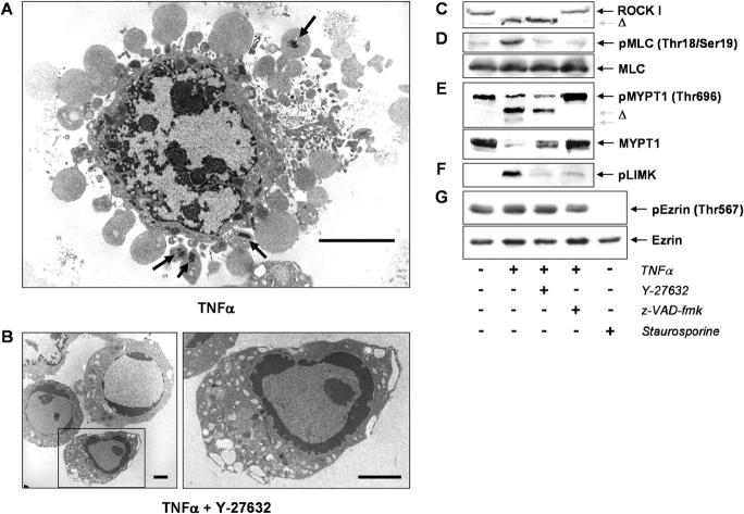
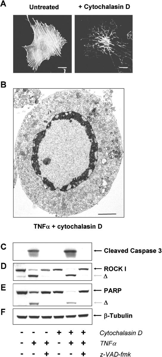
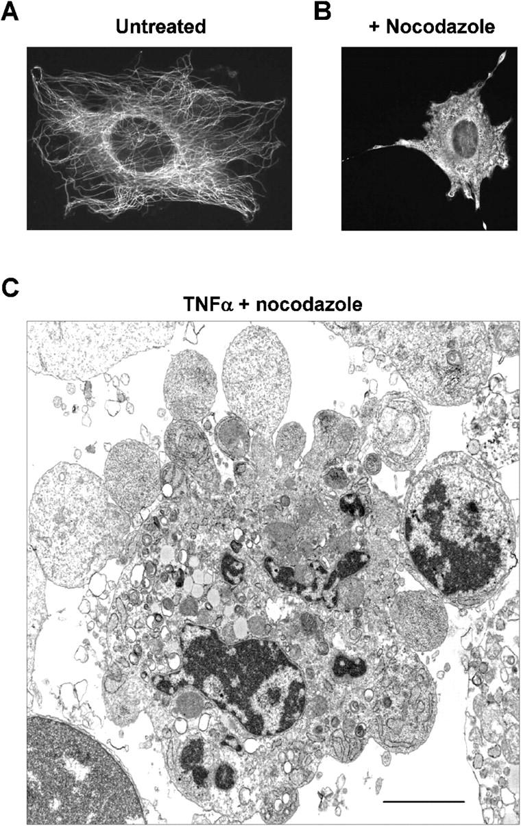
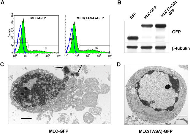
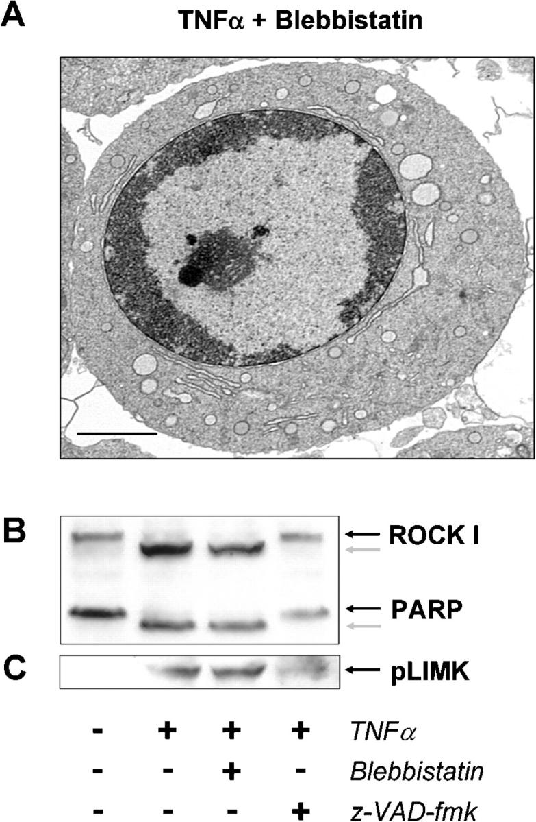
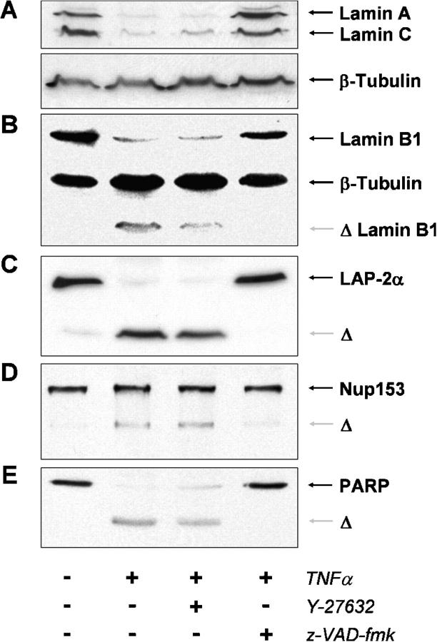
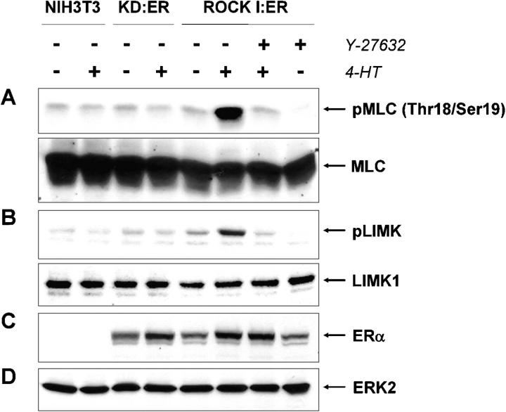
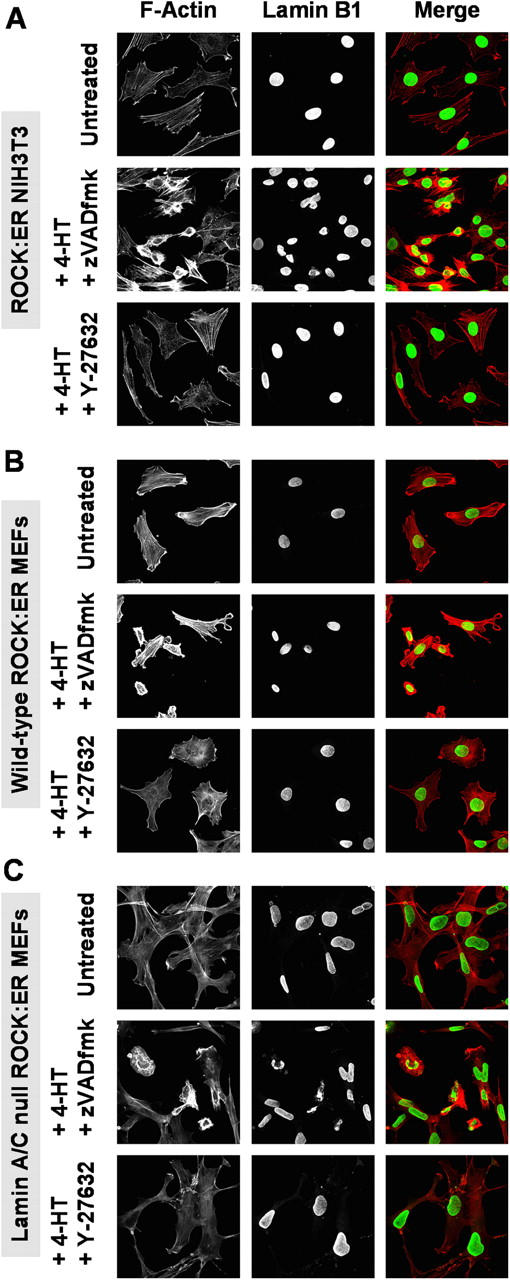
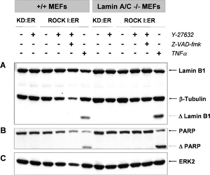
Membrane blebbing during the apoptotic execution phase results from caspase-mediated cleavage and activation of ROCK I. Here, we show that ROCK activity, myosin light chain (MLC) phosphorylation, MLC ATPase activity, and an intact actin cytoskeleton, but not microtubular cytoskeleton, are required for disruption of nuclear integrity during apoptosis. Inhibition of ROCK or MLC ATPase activity, which protect apoptotic nuclear integrity, does not affect caspase-mediated degradation of nuclear proteins such as lamins A, B1, or C. The conditional activation of ROCK I was sufficient to tear apart nuclei in lamin A/C null fibroblasts, but not in wild-type fibroblasts. Thus, apoptotic nuclear disintegration requires actin-myosin contractile force and lamin proteolysis, making apoptosis analogous to, but distinct from, mitosis where nuclear disintegration results from microtubule-based forces and from lamin phosphorylation and depolymerization.


Figures

Figure 1.
Figure 1.
Apoptotic nuclear breakdown is blocked following ROCK inhibition. (A) Transmission electron micrograph of an NIH 3T3 fibroblast treated with 25 ng/ml TNFα plus 10 μg/ml cycloheximide (CHX) for 2 h to induce apoptosis, showing large plasma membrane blebs, nuclear disintegration, and heterochromatin within blebs (arrows). Bar, 5 μm. (B) TEM of cells treated with 10 μM Y-27632 to block ROCK activity along with TNFα/CHX showing an absence of blebs and preservation of nuclear envelope integrity. Inset in left panel shown in right panel. Bars, 2 μm. (C–G) Serum-starved NIH 3T3 fibroblasts were left untreated or treated with 25 ng/ml TNFα plus 10 μg/ml CHX for 2 h to induce apoptosis, either in the absence or presence of 10 μM Y-27632 or 50 μM z-VAD-fmk as indicated. Lysates were prepared by direct lysis in 1× Laemmli sample buffer and samples run on 10% SDS-PAGE. Western blotting with antibodies against ROCK I (C), myosin light chain (MLC; D, bottom), and dually phosphorylated Thr18 Ser19 MLC (D, top), myosin binding subunit of the MLC phosphatase (MYPT1; E, bottom), and phosphorylated Thr696 MYPT1 (E, top), the common epitope of phospho-LIMK1 (Thr508; F) and phospho-LIMK2 (Thr505; F), and Ezrin (G, bottom) and phosphorylated Ezrin (Thr567; G, top). Phosphorylation of MLC and LIMK1/2 in apoptotic cells was blocked by ROCK inhibitor Y-27632 and by preventing caspase-mediated ROCK I cleavage with z-VAD-fmk. No differences in MYPT1 or Ezrin phosphorylation were evident, although MYPT1 was cleaved in a z-VAD-fmk–sensitive manner in apoptotic cells, removing ∼80 amino acids from the amino-terminal end.
Figure 2.
Figure 2.
Apoptotic nuclear breakdown is blocked following actin filament disruption. (A) Actin filaments were apparent in untreated NIH 3T3 cells (left), but cell morphology and actin filaments were profoundly affected by treatment with 2 μM cytochalasin D (right). Bars, 20 μm. (B) TEM of cell pretreated with 2 μM cytochalasin D to disrupt actin structures before induction of apoptosis, showing intact nucleus. Bar, 2 μm. (C) Activation of Caspase 3 and cleavage of ROCK I (D) and PARP (E) were not affected by cytochalasin treatment and disruption of the actin cytoskeleton before the induction of apoptosis. (F) Blotting for β-tubulin indicates equal loading across lanes.
Figure 3.
Figure 3.
Disruption of microtubular structures does not affect apoptotic nuclear breakdown. Microtubular structures were visualized with antibody against anti–β-tubulin antibody in untreated (A) or nocodazole (1 μM; B)-treated NIH 3T3 cells. (C) TEM of cell pretreated with 1 μM nocodazole to disrupt microtubules, as in B, before induction of apoptosis, showing marked blebbing and nuclear disintegration. Bar, 2 μm.
Figure 4.
Figure 4.
MLC phosphorylation is required for apoptotic nuclear disintegration. NIH 3T3 fibroblasts were transfected with a plasmid encoding GFP, myosin light chain fused to GFP (MLC-GFP), or MLC doubly mutated to Ala at the phosphorylation sites Thr18 Ser19 (MLC(TASA)-GFP). After allowing 24 h for protein expression, cells were sorted for GFP expression. (A) Autofluorescence of untransfected cells is indicated as R4, sorting was gated such that only GFP-positive cells with fluorescence intensity greater than all cells in R4 were collected (indicated as R3 in MLC-GFP and MLC(TASA)-GFP sorting profiles; GFP profile not depicted). Post-sorting analysis indicated that sorted cells were 99% GFP-positive. (B) Lysates prepared from equal numbers of GFP-positive cells were Western blotted for GFP to confirm equal protein expression (top) and for β-tubulin to confirm equal protein loading (bottom). (C) Cells were plated in medium containing 10% FCS for 5 h, and then placed in serum-free medium for 18 h. Apoptosis was induced with 25 ng/ml TNFα plus 10 μg/ml CHX for 2 h, and then nonadherent apoptotic cells were collected, fixed, and processed for TEM. Expression of GFP (not depicted) or MLC-GFP did not affect nuclear disintegration in apoptotic cells (arrows indicate heterochromatin in blebs). Bar, 2 μm. (D) Expression of nonphosphorylatable MLC(TASA)-GFP inhibited plasma membrane blebbing and nuclear fragmentation, although typical apoptotic chromatin condensation was evident. Bar, 2 μm.
Figure 5.
Figure 5.
Inhibition of myosin ATPase activity protects apoptotic nuclear integrity. (A) NIH 3T3 cells were coadministered blebbistatin (50 μM) along with the TNFα plus CHX apoptotic stimulus for 2 h before collection and processing for TEM. Bar, 2 μm. NIH 3T3 fibroblasts were untreated or treated with 50 μM blebbistatin to inhibit myosin ATPase activity or 50 μM z-VAD-fmk to inhibit caspase activity as indicated. Apoptosis was induced with TNFα/CHX where indicated. Lysates were prepared and Western blotted with antibodies against ROCK I and PARP (B) and phospho-LIMK1 and 2 (C). Blebbistatin affects apoptotic nuclear morphology without inhibiting caspase-mediated proteolysis or ROCK activity.
Figure 6.
Figure 6.
Degradation of nuclear lamins A/C and B1, LAP2α, Nup153, and PARP during apoptosis is unaffected by ROCK inhibition. NIH 3T3 fibroblasts were left untreated or treated with 10 μM Y-27632 to inhibit ROCK activity or 50 μM z-VAD-fmk to inhibit caspase activity as indicated. Apoptosis was induced with TNFα/CHX where indicated. Lysates were prepared and Western blotted with antibodies against lamins A and C (A), lamin B1 (B), LAP-2α (C), Nup-153 (D), and PARP (E). Equal loading was verified with an antibody against β-tubulin.
Figure 7.
Figure 7.
Conditionally active ROCK I:ER induces MLC and LIMK1 and 2 phosphorylation when stimulated with 4-HT. NIH 3T3 cells were left untreated or transduced with retrovirus encoding the conditionally active ROCK I:ER or kinase-dead KD:ER fusion proteins as indicated. After selection, stable pools were left untreated or treated with 1 μM 4-hydroxytamoxifen (4-HT) for 18 h as indicated, either in the absence or presence of ROCK inhibitor Y-27632 (10 μM). The phosphorylation status was examined with antibodies against MLC (A; dual Thr18/Ser19 phosphorylation, top panel; total MLC, bottom panel) and LIMK1 and 2 (B; LIMK1 Thr508, LIMK2 Thr505 phosphorylation, top panel; total LIMK 1, bottom panel). (C) Expression of KD:ER and ROCK I:ER confirmed by blotting with ERα antibody. (D) Equal protein levels confirmed by blotting with ERK2 antibody.
Figure 8.
Figure 8.
Active ROCK I induces contraction and membrane blebbing, but nuclear disruption only in lamin A/C null cells. Activation of ROCK I:ER with 4-HT resulted in increased filamentous actin structures and profound morphological responses. (A) ROCK I:ER-expressing NIH 3T3 cells had increased actin structures accompanied by cell contraction and membrane blebbing in ∼50% of cells after treatment with 1 μM 4-HT, either in the presence (middle panels) or absence (not depicted) of z-VAD-fmk. Coadministration of ROCK inhibitor Y-27632 blocked the effects on actin structures and morphological responses (bottom panels). Nuclear morphology and integrity was assessed by staining for lamin B1 (middle column), ROCK-induced contraction and blebbing also resulted in contracted and distorted nuclei that remained intact (middle row), which could be blocked by ROCK inhibitor Y-27632 (bottom row). (B) Primary wild-type mouse embryo fibroblasts (MEFs) transduced with ROCK I:ER responded to 4-HT treatment with increased filamentous actin structures and many contracted, blebbing cells (middle row), responses that could be blocked by ROCK inhibitor Y-27632 (bottom row). ROCK-induced contraction and blebbing also resulted in contraction of nuclei (middle column) as determined by lamin B1 staining. (C) Primary lamin A/C null MEFs transduced with ROCK I:ER also responded to 4-HT with increased filamentous actin structures, cell contraction, and membrane blebbing (middle row), which could be blocked with ROCK inhibitor Y-27632. Nuclei (middle column) in contracted blebbing cells were severely distorted and often disrupted, even in the presence of z-VAD-fmk (middle row), which could be blocked by ROCK inhibitor Y-27632.
Figure 9.
Figure 9.
ROCK I does not activate caspases. Wild-type and lamin A/C null MEFs expressing ROCK I:ER or kinase dead KD:ER were left untreated or treated with 1 μM 4-HT, either in the absence or presence of z-VAD-fmk as indicated. As a positive control for caspase activation, cells were treated with 25 ng/ml TNFα plus 10 μg/ml CHX. Western blotting revealed that 4-HT treatment of KD:ER- or ROCK I:ER-expressing wild-type and lamin A/C null fibroblasts did not induce cleavage of lamin B1 (A) or PARP (B) although each was cleaved in response to TNFα treatment. (C) Western blotting for ERK2 reflects protein levels across samples.

References

    1. Amano, T., K. Tanabe, T. Eto, S. Narumiya, and K. Mizuno. 2001. LIM-kinase 2 induces formation of stress fibres, focal adhesions and membrane blebs, dependent on its activation by Rho-associated kinase-catalysed phosphorylation at threonine-505. Biochem. J. 354:149–159. - PMC - PubMed
    1. Apel, E.D., R.M. Lewis, R.M. Grady, and J.R. Sanes. 2000. Syne-1, a dystrophin- and Klarsicht-related protein associated with synaptic nuclei at the neuromuscular junction. J. Biol. Chem. 275:31986–31995. - PubMed
    1. Beaudouin, J., D. Gerlich, N. Daigle, R. Eils, and J. Ellenberg. 2002. Nuclear envelope breakdown proceeds by microtubule-induced tearing of the lamina. Cell. 108:83–96. - PubMed
    1. Broers, J.L., N.M. Bronnenberg, H.J. Kuijpers, B. Schutte, C.J. Hutchison, and F.C. Ramaekers. 2002. Partial cleavage of A-type lamins concurs with their total disintegration from the nuclear lamina during apoptosis. Eur. J. Cell Biol. 81:677–691. - PubMed
    1. Bubb, M.R., I. Spector, A.D. Bershadsky, and E.D. Korn. 1995. Swinholide A is a microfilament disrupting marine toxin that stabilizes actin dimers and severs actin filaments. J. Biol. Chem. 270:3463–3466. - PubMed

Publication types

MeSH terms

Substances