. 2005 Feb;25(3):888-95.
doi: 10.1128/MCB.25.3.888-895.2005.

Mechanisms of transcription-replication collisions in bacteria

Affiliations

Mechanisms of transcription-replication collisions in bacteria

Ekaterina V Mirkin et al. Mol Cell Biol. 2005 Feb.

Abstract

While collisions between replication and transcription in bacteria are deemed inevitable, the fine details of the interplay between the two machineries are poorly understood. In this study, we evaluate the effects of transcription on the replication fork progression in vivo, by using electrophoresis analysis of replication intermediates. Studying Escherichia coli plasmids, which carry constitutive or inducible promoters in different orientations relative to the replication origin, we show that the mutual orientation of the two processes determines their mode of interaction. Replication elongation appears not to be affected by transcription proceeding in the codirectional orientation. Head-on transcription, by contrast, leads to severe inhibition of the replication fork progression. Furthermore, we evaluate the mechanism of this inhibition by limiting the area of direct contact between the two machineries. We observe that replication pausing zones coincide exactly with transcribed DNA segments. We conclude, therefore, that the replication fork is most likely attenuated upon direct physical interaction with the head-on transcription machinery.


Figures

FIG. 1.
FIG. 1.
Schematic representation of the head-on and codirectional collisions between replication and transcription. The replication fork (left) and RNA polymerase (right) are shown with leading- and lagging-strand DNA polymerases as ovals and DNA helicase DnaB as a hexagon. Solid lines, DNA strands; broken lines, RNA strands.
FIG. 2.
FIG. 2.
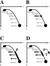
Schematic representation of the two-dimensional electrophoresis analysis of replication intermediates. (A) Each dot on the bubble arc represents a replication intermediate, starting from the small bubble at the origin of replication (bottom) and going upward to the biggest, fully replicated bubble (top). The smooth bubble arc reflects the uniform speed of replication throughout the plasmid. (B) If replication progression is slowed down at a particular spot in the plasmid (bold replication intermediate), a distinct bulge appears on the arc. (C) Transcription from the head-on-oriented promoter (P) can inhibit replication progression in the whole DNA segment, separating the promoter and the origin. (D) Insertion of the transcription terminator (T) limits the area of replication inhibition.
FIG. 3.
FIG. 3.
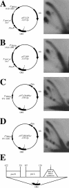
Replication inhibition by the head-on-oriented promoter for the seven tRNAs. In plasmids pP7-CD (A) and pP7-CDΔPlac (C), the P7 promoter faces the direction of replication. In the plasmids pP7-HO (B) and pP7-HOΔPlac (D), the P7 promoter faces replication head-on. The structure of the plasmid replication origin is shown (E). In all of the plasmids, nucleotide position 1 corresponds to the replication start site. Positions of the P7 promoter relative to the replication start site in each plasmid are indicated. Replication is inhibited when transcription faces replication head-on.
FIG. 4.
FIG. 4.
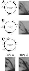
Active transcription is required for the replication inhibition. In the plasmid pTrc-CDΔPlac, the trc promoter faces the direction of replication (A). In the plasmid pTrc-HOΔPlac, the trc promoter faces replication head-on (B). Replication of the pTrc-HO/LacIq plasmid in the absence or presence of IPTG when the promoter is off or on, respectively, is shown in panel C. In all plasmids, nucleotide position 1 corresponds to the replication start site. Positions of the trc promoter relative to the replication start site in each plasmid are indicated. Replication inhibition is evident when transcription is on and faces replication head-on.
FIG. 5.
FIG. 5.
Insertion of transcription termination signals limits the area of replication inhibition. Replication of the plasmids, where transcription from the head-on trc promoter is limited to either 200- (A) or 400-bp (B) DNA segments, in the absence or presence of IPTG, is shown. In these plasmids, nucleotide position 1 corresponds to the replication start site. Positions of the trc promoter and transcription terminators relative to the replication start site are indicated.
FIG. 6.
FIG. 6.
Mapping of the replication stop zones. (A) Schematic representation of the in-gel digest of the replication intermediates after the first dimension of the two-dimensional gel electrophoresis. The vertical lines show the positions of the restriction sites immediately upstream (PstI) and downstream (HindIII) of the transcribed area, situated between the trc promoter (P) and transcription terminator (T). Specifically, the HindIII site is located in nucleotide position 1708 relative to the replication start site in both plasmids, while the PstI site is located in position 2280 in the pTrc-HO/T1T2-200 plasmid and in the position 2483 in the pTrc-HO/T1T2-400 plasmid. Replication intermediates are shown as bubbles, and those drawn with thick lines reflect replication stalling. Upon HindIII digestion, stalled intermediates become Y shaped, while after PstI digestion they remain bubble shaped. Replication intermediates of plasmids pTrc-HO/T1T2-200 and pTrc-HO/T1T2-400, isolated upon growth in the presence of IPTG, were digested with either HindIII (B) or PstI (C) after the first dimension of the electrophoresis. Note the arc-to-line transition of stalled replication intermediates, shown by thick arrows, in panel B but not in panel C. The partially digested underreplicated intermediates, migrating between the arc and the line in panel B, are shown by thin arrows (see text for details).

Similar articles

Cited by

References

    1. Aman, E., B. Ochs, and K.-J. Abel. 1988. Tightly regulated tac promoter vectors useful for the expression of unfused and fused proteins in Escherichia coli. Gene 69:301-315. - PubMed
    1. Blattner, F. R., G. Plunkett III, C. A. Bloch, N. T. Perna, V. Burland, M. Riley, J. Collado-Vides, J. D. Glasner, C. K. Rode, G. F. Mayhew, J. Gregor, N. W. Davis, H. A. Kirkpatrick, M. A. Goeden, D. J. Rose, B. Mau, and Y. Shao. 1997. The complete genome sequence of Escherichia coli K-12. Science 277:1453-1474. - PubMed
    1. Brewer, B. J. 1988. When polymerases collide: replication and the transcriptional organization of the E. coli chromosome. Cell 53:679-686. - PubMed
    1. Brewer, B. J., and W. L. Fangman. 1994. Initiation preference at a yeast origin of replication. Proc. Natl. Acad. Sci. USA 91:3418-3422. - PMC - PubMed
    1. Brewer, B. J., and W. L. Fangman. 1987. The localization of replication origins on ARS plasmids in S. cerevisiae. Cell 51:463-471. - PubMed

Publication types

MeSH terms

Substances

LinkOut - more resources