. 2005 Feb;25(3):969-78.
doi: 10.1128/MCB.25.3.969-978.2005.

Desmosomal cadherin misexpression alters beta-catenin stability and epidermal differentiation

Affiliations

Desmosomal cadherin misexpression alters beta-catenin stability and epidermal differentiation

Matthew J Hardman et al. Mol Cell Biol. 2005 Feb.

Abstract

Desmosomal adhesion is important for the integrity and protective barrier function of the epidermis and is disregulated during carcinogenesis. Strong adhesion between keratinocytes is conferred by the desmosomal cadherins, desmocollin (Dsc) and desmoglein. These constitute two gene families, members of which are differentially expressed in epidermal strata. It has been suggested that this stratum-specific expression regulates keratinocyte differentiation. We tested this hypothesis by misdirecting the expression of the basally abundant desmosomal cadherins Dsc3a and Dsc3b to suprabasal differentiating keratinocytes in transgenic mice. No phenotype was apparent until adulthood, when mice developed variable ventral alopecia and had altered keratinocyte differentiation within affected areas. The follicular changes were reminiscent of changes in transgenic mice with an altered beta-catenin stability. Stabilized beta-catenin and increased beta-catenin transcriptional activity were demonstrated in transgenic mice prior to the phenotypic change and in transgenic keratinocytes as a consequence of transgene expression. Hence, a link between desmosomal cadherins and beta-catenin stability and signaling was demonstrated, and it was shown that desmocollin cadherin expression can affect keratinocyte differentiation. Furthermore, the first function for a "b-type" desmocollin cadherin was demonstrated.


Figures
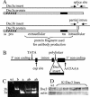
FIG. 1.
FIG. 1.
(A) Dsc3a and -3b cDNA inserts showing the site of the alternatively spliced C-terminal intron (asterisk) and the partial intron sequence in the Dsc3b construct. The unspliced b isoform terminates within the intron. Arrows show primer sequences used for isoform-specific transgene detection. Below the inserts, boxes show encoded proteins with signal sequences (ss), “pro” sequences (hatched), extracellular domains, transmembrane domains (tm), intracellular domains, and regions used for antibody production. (B) Keratin 1 targeting vector (14, 35) showing Dcs3 insert site. (C) PCR analysis of genomic DNA. wt, wild-type DNA showing absence of amplification; b, Dsc3b transgenic; a, Dsc3a transgenic; ab, Dsc3a/b double transgenic showing isoform-specific transgene detection. (D) Western analysis of Dsc3 showed only minor differences between wild-type and transgenic protein levels in newborn Dsc3a lines.
FIG. 2.
FIG. 2.
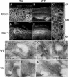
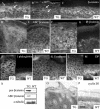
Desmocollin distribution in K1Dsc3 transgenic affected skin. (A) Immunohistochemical detection of Dsc3 protein in transgenic skin showing Dsc3 presence in all strata and a loss of the graded basally abundant expression pattern characteristic of control skin (footpad skin is used to show graded expression in the control [B]). (C) Higher magnification showing Dsc3 punctate membranous expression in basal and suprabasal layers of transgenic skin, indicating desmosomal incorporation. Desmocollin 1 protein distribution appears unaffected in transgenic (D) and littermate control (E) animals, in which it shows reciprocal graded expression compared to Dsc3, with peak expression in the granular layer (compare panels B and E). (F to K) Immunogold detection of Dsc3 in individual desmosomes from distinct epidermal strata in transgenic and control mice. In the wild-type epidermis, Dsc3 is incorporated in desmosomes in lower (F) and middle (G) strata but is almost undetectable in upper strata (H). In contrast, the Dsc3 protein was readily detected in upper strata in the K1Dsc3 epidermis (K), demonstrating that transgene-encoded Dsc3 incorporates into desmosomes. WT, wild type; TG, transgenic; gr, granular layer; sbl, suprabasal layer; bl, basal layer; de, dermis. Bar, 40 μm (A, B, D, and E), 15 μm (C), or 125 nm (F to K).
FIG. 3.
FIG. 3.
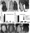
Hair defect in K1Dsc3 transgenic mice. (A to C) Transgenic mice developed a progressive ventral alopecia by 10 to 12 weeks which was absent in control littermates (B and C [compare transgenics with control littermates on the right]). The extent of ventral alopecia varied from a small ventral patch (A) to extensive ventral hair loss (C). Hairs in the affected regions were sparse and consisted entirely of zigzag hairs (D), while adjacent unaffected regions contained all four hair types in predicted ratios (E) (6). (F to I) Ventral alopecia accompanies the first postnatal hair cycle. The depilation of ventral skin 2 weeks after birth (F and H) resulted in phenotypic alopecia by 9 days in transgenic skin (G), while wild-type skin recovered (I).
FIG. 4.
FIG. 4.
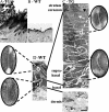
K1Dsc3 transgenic mouse epidermis from ventral skin (A) was substantially thickened compared to control epidermis from the same site (B). The K1Dsc3 epidermis also showed significant hyperkeratosis (A). (C) Ultrastructural analysis by electron microscopy showed that the thicker epidermis was due to a substantially increased number of spinous layers (acanthosis) and granular layers (hypergranulosis) in the transgenic epidermis compared to the wild type (D). In addition, the flattened, anucleate keratinocytes of the transgenic stratum corneum (C, black arrow) appeared less compact than control keratinocytes (D, black arrow), and keratohyalin granules were abnormally large (C, white arrow). Desmosomes from all layers of transgenic skin (C, insets) were ultrastructurally comparable to those of control skin (D, insets). Bar, 100 μm (A and B), 5 μm (C and D), or 100 nm (insets).
FIG. 5.
FIG. 5.
Altered terminal differentiation in transgenic epidermis overexpressing Dsc3 suprabasally. (A, C, E, G, I, K, and M) Epidermis from transgenic ventral skin; (B, D, F, H, J, L, and N) Site-matched epidermis from littermate controls. (A and B) Ki67 immunohistochemistry showed that basal layer proliferation was increased in K1Dsc3 epidermis and that proliferative cells were found suprabasally. Arrows show proliferating cells in control epidermis. (C and D) K14immunohistochemistry showed broadened basal and suprabasal expression of keratin 14 in transgenic skin (C). K14 is usually confined to the basal layer in wild-type skin. The inset in panel D shows basal K14 expression in the thicker epidermis from the foot of a control animal, as basal expression is difficult to demonstrate in the thin ventral skin of wild-type mice. (E and F) K1 immunohistochemistry showed broadened expression of suprabasally expressed keratin 1, which was associated with acanthosis in the transgenic epidermis (E). The inset in panel F shows suprabasal K1 expression in the thicker epidermis from the foot of a control animal. Involucrin (G and H), loricrin (I and J), and filaggrin (K and L) also showed broadened expression patterns in the K1Dsc3 epidermis (G, I, and K) which were associated with hypergranulosis compared to control epidermis (H, J, and L), although the differentiation patterns were essentially normal. Keratin 6 was induced in the thickened epidermis of transgenic mice (M) but was undetectable in the control interfollicular epidermis (N). Note the normal follicular expression. Bar, 30 μm (A, B, and G to N), 40 μm (C to F), or 80 μm (insets).
FIG. 6.
FIG. 6.
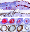
Altered lipid distribution, follicle degeneration, and associated de- or transdifferentiation of follicles in K1Dsc3 transgenic skin. (A) SSLF was reduced or entirely absent in transgenic skin, as visualized by oil red O staining, compared to the prominent SSLF in site-matched control littermates (B). Also, the follicles were degenerated (A, arrow) and sebaceous glands were abnormally distributed and enlarged in transgenic skin (A) compared to control skin (B, arrow shows a normal cross-sectioned follicle). (C and D) In the transgenic, K1, which is normally expressed in the interfollicular epidermis (IFE) and the uppermost part of the follicle (D, arrow), was expressed in utriculi and dermal cysts deep in the dermis (C, arrow to utriculus; DC, dermal cyst). (E) K1Dsc3 skin showed a range of dermal cyst-like structures, ranging from sebum filled (left) to highly keratinized (right). (E [left], F, and G) Serial sections stained with oil red O, K14, and K1, respectively. This sebum-filled (E) cyst expressed both K14 (F) and, unexpectedly, the exclusively IFE marker K1 (G). (H and I) Cysts deep in the dermis stained with K1 (H) and loricrin (I) markers, which are not expressed in follicles but are characteristic of a differentiated IFE. Bar, 150 μm (A to D) or 40 μm (E to I).
FIG. 7.
FIG. 7.
Changed β-catenin abundance and distribution in K1Dsc3 transgenic skin. (A and B) A pan-β-catenin antibody demonstrated membranous β-catenin associated with adherens junctions in all living strata of transgenic (A, B) and wild-type (C) skin. However, β-catenin was also detectable cytoplasmically and in nuclei (A and B, arrows) in suprabasal layers of transgenic skin. (D and E) The altered β-catenin pattern in transgenic skin was due to increased cytoplasmic and nuclear active β-catenin (ABC, detected with an antibody to an unphosphorylated N-terminal β-catenin epitope [42]) in the transgenic epidermis (D) compared to the control (E). (F and G) Transgenic skin had substantially increased cytoplasmic and nuclear inactive β-catenin (detected with anti-phospho-β-catenin, an antibody to a phosphorylated N-terminal β-catenin epitope [37]) in all layers of the transgenic epidermis (F). Labeling was confined to the cytoplasm of basal cells in the control epidermis (G). Note the perinuclear staining of keratinocytes in transgenic skin (G). In contrast, other protein components of the adherens junction (H and I, plakoglobin; J and K, E-cadherin; L and M, desmoplakin [DP]) showed similar distributions in transgenic (H, J, and L) and control (I, K, and M) epidermis. (N) Enhanced levels of total (pan) and active β-catenin in K1Dsc3 epidermis were confirmed by Western analysis. (O) Cyclin D immunolocalized to nuclei of suprabasal as well as basal strata in K1Dsc3a transgenic skin but was confined to the basal layer in wild-type epidermis (M [footpad skin was used to provide a similar number of suprabasal layers as the hyperproliferative transgenic control]). Bar, 20 μm (A to G), 30 μm (H to M), or 80 μm (O and P).
FIG. 8.
FIG. 8.
(A) Primary keratinocytes from K1Dsc3a newborn epidermis (TG) show enhanced ability to activate the TOP-flash reporter gene compared to control keratinocytes (WT) or keratinocytes transfected with constitutively active β-catenin (βcat), indicating an enhanced ability to activate Lef/Tcf downstream genes. (B) A human keratinocyte cell line (N/TERT-1) transfected with K1Dsc3a and K1Dsc3b transgenes did not activate ΤΟP-flash in the presence of 0.4 mM calcium, a level in which desmosomes can form but expression of the transgene promoter is minimal. However, upon transfer to a high calcium level (1.5 mM) to activate the transgene promoter, K1Dsc3a and K1Dsc3b both activated TOP-flash activity in a dose-dependent manner compared to cells transfected with TOP-flash alone (TOP), FOP-flash alone (FOP), or a transgene and FOP-flash (not shown). Cells transfected with constitutively active β-catenin/TOP-flash (βcat) provided a positive control. Note the higher endogenous β-catenin activity in keratinocytes transfected with TOP-flash alone and grown in low (0.4 mM) calcium than in the same keratinocytes grown in 1.5 mM calcium. Error bars, standard errors of the means.

Similar articles

Cited by

References

    1. Abdalkhani, A., R. Sellers, J. Gent, H. Wulitich, S. Childress, B. Stein, R. E. Boissy, J. J. Wysolmerski, J. Foley, P. Dann, J. Hong, J. Cosgrove, B. Dreyer, D. Rimm, M. Dunbar, W. Philbrick, and J. Wysolmerski. 2002. Nipple connective tissue and its development: insights from the K14-PTHrP mouse. Mech. Dev. 115:63-77. - PubMed
    1. Bonne, S., B. Gilbert, M. Hatzfeld, X. Chen, K. J. Green, and F. van Roy. 2003. Defining desmosomal plakophilin-3 interactions. J. Cell Biol. 161:403-416. - PMC - PubMed
    1. Chen, X., S. Bonne, M. Hatzfeld, F. van Roy, and K. J. Green. 2002. Protein binding and functional characterization of plakophilin 2. Evidence for its diverse roles in desmosomes and beta-catenin signaling. J. Biol. Chem. 277:10512-10522. - PubMed
    1. Cheng, X., K. Mihindukulasuriya, Z. Den, A. P. Kowalczyk, C. C. Calkins, A. Ishiko, A. Shimizu, and P. J. Koch. 2004. Assessment of splice variant-specific functions of desmocollin 1 in the skin. Mol. Cell. Biol. 24:154-163. - PMC - PubMed
    1. Dickson, M. A., W. C. Hahn, Y. Ino, V. Ronfard, J. Y. Wu, R. A. Weinberg, D. N. Louis, F. P. Li, and J. G. Rheinwald. 2000. Human keratinocytes that express hTERT and also bypass a p16(INK4a)-enforced mechanism that limits life span become immortal yet retain normal growth and differentiation characteristics. Mol. Cell. Biol. 20:1436-1447. - PMC - PubMed

Publication types

MeSH terms

LinkOut - more resources