. 2005 Feb;25(3):1041-53.
doi: 10.1128/MCB.25.3.1041-1053.2005.

Silencing of unpaired chromatin and histone H2A ubiquitination in mammalian meiosis

Affiliations

Silencing of unpaired chromatin and histone H2A ubiquitination in mammalian meiosis

Willy M Baarends et al. Mol Cell Biol. 2005 Feb.

Abstract

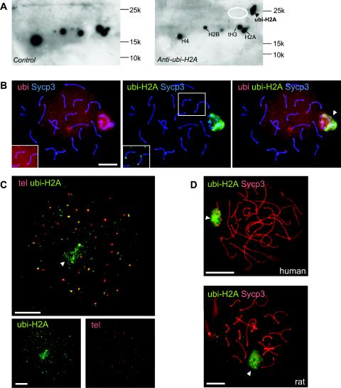
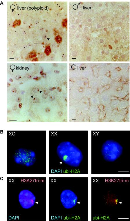
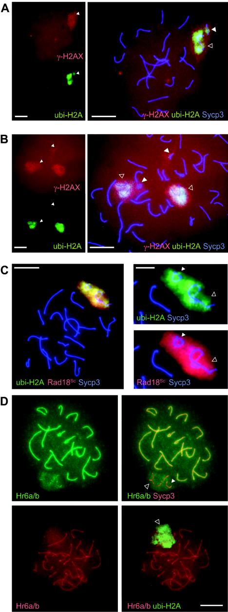
During meiotic prophase in male mammals, the X and Y chromosomes are incorporated in the XY body. This heterochromatic body is transcriptionally silenced and marked by increased ubiquitination of histone H2A. This led us to investigate the relationship between histone H2A ubiquitination and chromatin silencing in more detail. First, we found that ubiquitinated H2A also marks the silenced X chromosome of the Barr body in female somatic cells. Next, we studied a possible relationship between H2A ubiquitination, chromatin silencing, and unpaired chromatin in meiotic prophase. The mouse models used carry an unpaired autosomal region in male meiosis or unpaired X and Y chromosomes in female meiosis. We show that ubiquitinated histone H2A is associated with transcriptional silencing of large chromatin regions. This silencing in mammalian meiotic prophase cells concerns unpaired chromatin regions and resembles a phenomenon described for the fungus Neurospora crassa and named meiotic silencing by unpaired DNA.


Figures

FIG. 1.
FIG. 1.
ubi-H2A marks the XY bodies and telomeres of pachytene spermatocytes. A: Western blots of basic nuclear proteins separated on two-dimensional AUT-SDS-polyacrylamide gels (first dimension, AUT; second dimension, SDS) were stained with anti-ubi-H2A (right panel) or with second antibody only (control, left panel). A specific signal is detected at the expected position for ubi-H2A (arrowhead). The approximate positions of histones H2A, H2B, tH3, and H4 are indicated for reference. Positions and molecular weights of marker proteins are indicated. The expected approximate position of ubi-H2B is indicated by the white ellipse, and the blots are overexposed to show that no ubi-H2B is detected. B: Spermatocyte spread nuclei from wild-type mice stained with a mouse monoclonal IgG antibody against Sycp3 (blue), a rabbit polyclonal antiubiquitin antibody (red), and a mouse monoclonal IgM antibody against ubi-H2A (green). Both ubi and ubi-H2A mark XY body chromatin (arrowhead in right panel, showing the merged signals). Anti-ubi also detects other ubiquitinated proteins associated with chromatin in the rest of the nucleus. Upon overexposure, ubi and ubi-H2A are detected at the ends of some synaptonemal complex axes (insets). C: FISH analysis of telomeres (tel) (red) followed by immunocytochemistry with anti-ubi-H2A (green) of an early diplotene cell. Diplotene is identified by the presence of a low level of ubi-H2A in the XY body (arrowhead) and the high number of telomeric foci (more than 40, with double foci still attached counted as one) and splitting of telomeres. The upper panel shows that around 30 telomere foci colocalize, at least partially, with ubi-H2A. A total of approximately 100 ubi-H2A foci are observed outside the XY body area, indicating that chromatin regions other than the XY body and (near) telomeric regions also accumulate ubi-H2A. The bottom panels show lower-magnification images of the single fluorescent signals. D: Spermatocyte spread nuclei from rat and human stained with anti-Sycp3 (red) and anti-ubi-H2A (green). ubi-H2A marks XY body chromatin (arrowhead) in both species. Bars, 20 μm.
FIG. 2.
FIG. 2.
ubi-H2A marks the inactive X chromosome (Barr body) in female somatic cells. A: Immunohistochemical analysis of ubi-H2A localization in rat female liver and kidney sections and in male liver sections. Control (C liver): incubation without first antibody. In female kidney nuclei, high-level accumulation of ubi-H2A is seen as a single nuclear spot (arrowhead). In addition, two spots are often observed in polyploid female liver nuclei (arrowhead). These spots are not observed in male tissues. Bars, 10 μm. B: Immunocytochemical detection of ubi-H2A in XO, XX, and XYtdym1 pregranulosa cells. A variable number of small foci, of unknown nature, is present in all nuclei, but only the XX pregranulosa cells accumulate ubi-H2A in a single large spot in the periphery of the nucleus (arrowhead). Bar, 20 μm. C: Immunocytochemical detection of H3 lysine 27 trimethylation (H3K27tri-m) (red) and ubi-H2A (green) in XX pregranulosa cells. H3 lysine 27 methylation and ubi-H2A largely colocalize in a single large spot in the periphery of the nucleus (arrowhead). Bar, 20 μm.
FIG. 3.
FIG. 3.
ubi-H2A marks the partially synapsed 113 translocation bivalent of T/T′ spermatocytes. The top row shows four T/T′ pachytene spermatocyte spread nuclei stained with anti-ubi-H2A (green) and anti-Sycp3 (red). The closed arrowheads indicate the location of the 113 bivalent, at different positions with respect to the XY body (open arrowheads). Bar, 20 μm. The images in the second row represent the four defined groups of 113 synapsis morphologies, detected with anti-Sycp3 staining and shown at a higher magnification (bar, 5 μm). PR, partially synapsed rest; PA, partially synapsed A shape; PH, partially synapsed horseshoe shape; CS, complete synapsis. The table shows that an increasing fraction of 113 bivalents is ubi-H2A negative when synapsis is more complete. Absolute numbers of scored nuclei with no ubi-H2A signal (−), a low ubi-H2A signal (+/−), or a clear ubi-H2A signal (+) are indicated. Tot, total number of nuclei counted.
FIG. 4.
FIG. 4.
Ubiquitination of H2A is not associated with increased trimethylation of histone 3 at lysine 9 or lysine 27. A: Double immunostaining of a T/T′ pachytene spermatocyte spread nucleus with anti-Sycp3 (red) and anti-RNA polymerase (pol) II (green). The inset shows a larger magnification of the area containing the XY body (open arrowhead) and the 113 bivalent (closed arrowhead). B: Triple immunostaining of a T/T′ pachytene spermatocyte spread nucleus with anti-trimethylated H3 lysine 9 (H3K9tri-m) (red), anti-ubi-H2A (green), and anti-Sycp3 (blue). The inset shows double staining of anti-trimethylated H3 lysine 9 and anti-Sycp3 in the area containing the XY body (open arrowhead) and the 113 bivalent (closed arrowhead). Both the translocation bivalent (PH group) and the XY body accumulate trimethylated H3 lysine 9 and ubi-H2A. The level of ubi-H2A appears to be higher in areas that contain a somewhat lower trimethylated H3 lysine 9 signal. C: As in panel B, but this late pachytene spermatocyte shows no accumulation of trimethylated H3 lysine 9 in the area containing the XY body and the 113 bivalent (PA group). D: As in panel A, but this spread nucleus was incubated with anti-trimethylated H3 lysine 27 (H3K27tri-m) (green) and anti-Sycp3 (red). H3 lysine 27 trimethylation is relatively low in the area that covers the XY body and the 113 bivalent (PR group). Bar, 20 μm; bar in inset, 10 μm.
FIG. 5.
FIG. 5.
ubi-H2A, γ-H2AX, and Rad18Sc colocalize in unsynapsed chromatin regions of late pachytene spermatocytes. A: Triple immunostaining of a T/T′ pachytene spermatocyte spread nucleus with anti-γ-H2AX (red), anti-ubi-H2A (green), and anti-Sycp3 (blue). The left panel shows single immunostaining (lower magnification), and the right panel shows triple immunostaining (higher magnification). Both γ-H2AX and ubi-H2A accumulate on the partially synapsed 113 bivalent (PR group). B: As in panel A, but two spread nuclei that accumulate γ-H2AX but not ubi-H2A on partially synapsed 113 bivalents (PA and PH group) are shown. C: The left panel shows triple immunostaining of a T/T′ pachytene spermatocyte spread nucleus with anti-Sycp3 (blue), anti-Rad18Sc (red), and anti-ubi-H2A (green). Bar, 20 μm. The right panels show a higher magnification (bar, 10 μm) of the ubi-H2A/Sycp3 and the Rad18Sc/Sycp3 double staining of an XY body (open arrowhead) and a PA group 113 bivalent (closed arrowhead). D: The upper two panels show a T/T′ spermatocyte spread nucleus immunostained with anti-Hr6a/b (green) and anti-Sycp3 (red). A diffuse Hr6a/b signal covers the XY body (open arrowhead) and the 113 bivalent (closed arrowhead). The highest Hr6a/b signal is observed on synapsed synaptonemal complex. The lower two panels show a T/T′ spermatocyte spread nucleus immunostained with anti-Hr6a/b (red) and anti-ubi-H2A (green). The diffuse Hr6a/b signal (most likely covering the XY body and the 113 bivalent [open arrowhead]) colocalizes with ubi-H2A. Bar, 20 μm.
FIG. 6.
FIG. 6.
ubi-H2A and Rad18Sc mark unsynapsed chromosome regions in oocytes. A: Two XY oocyte spread nuclei immunostained with anti-Rad18Sc (green) and anti-Sycp3 (red). The locations of the X and Y chromosomes are indicated. The smaller panels to the right of each nucleus show enlargements of the X and Y chromosomal areas with the white FISH signals for either X or Y (arrows). In the nucleus shown on the left, X and Y are partially synapsed and Rad18Sc accumulates mainly on X-chromosomal chromatin. In the nucleus shown on the right, X and Y have separated, and Rad18Sc accumulation is observed only on the Y chromosome. In this nucleus, the X-chromosomal Sycp3 axis is short and thick, indicating self-synapsis. Note that some X FISH signal is still visible after Y FISH (see Materials and Methods). B: Two XY oocyte spread nuclei immunostained with anti-ubi-H2A (green) and anti-Sycp3 (red). The smaller panels to the right of each nucleus show enlargements of the X and Y chromosomal areas with the white FISH signals for either X or Y (arrows). In both nuclei, X and Y have separated, but ubi-H2A marks the X chromosome in the nucleus in the left panel (the Y chromosome appears to undergo heterologous synapsis) and the Y chromosome in the nucleus in the right panel (the X chromosome appears to be unsynapsed). C: XX oocyte spread nucleus immunostained with anti-Sycp3 (red) and either anti-ubi-H2A (left panel) or anti-Rad18Sc (right panel) (green). The two images in the middle show enlargements (bar, 10 μm) of the aberrant synaptonemal complex morphology (Sycp3 staining) in the respective ubi-H2A (1)- and Rad18Sc (2)-positive regions. Bars, 20 μm unless otherwise indicated.
FIG. 7.
FIG. 7.
Transcriptional repression of XY chromosomes in XO and XYtdym1 oocytes. A: Early (ep) and late (lp) pachytene XYtdym1 oocyte spread nuclei immunostained with anti-Sycp3 (red) and anti-RNA polymerase (pol) II (green). The area with unsynapsed synaptonemal complex axes and low RNA pol II immunoexpression is circled. B: Pachytene XO oocyte spread nucleus with accumulation of γ-H2AX (red) and ubi-H2A (green) on the unsynapsed X chromosome (arrowhead). Immunostaining of Sycp3 is shown in blue. The middle panel shows the region that contains the unsynapsed X chromosome. The panels on the right show the single γ-H2AX (red) and ubi-H2A (green) immunostainings. C: The four panels show one XO oocyte spread nucleus. RNA pol II immunoexpression (green) is relatively low in regions with intense DAPI staining (blue) and also in a Rad18Sc-positive region (red, Rad18) that is circled in the merge and DAPI panels. Bars, 20 μm.

References

    1. Baarends, W. M., J. W. Hoogerbrugge, H. P. Roest, M. Ooms, J. Vreeburg, J. H. J. Hoeijmakers, and J. A. Grootegoed. 1999. Histone ubiquitination and chromatin remodeling in mouse spermatogenesis. Dev. Biol. 207:322-333. - PubMed
    1. Bailly, V., J. Lamb, P. Sung, S. Prakash, and L. Prakash. 1994. Specific complex formation between yeast RAD6 and RAD18 proteins: a potential mechanism for targeting RAD6 ubiquitin-conjugating activity to DNA damage sites. Genes Dev. 8:811-820. - PubMed
    1. Barr, M. L., and E. G. Bertram. 1949. A morphological distinction between neurones of the male and female, and the behaviour of the nucleolar satellite during accelerated nucleoprotein synthesis. Nature 163:676-677. - PubMed
    1. Bean, C. J., C. E. Schaner, and W. G. Kelly. 2004. Meiotic pairing and imprinted X chromatin assembly in Caenorhabditis elegans. Nat. Genet. 36:100-105. - PMC - PubMed
    1. Briggs, S. D., T. Xiao, Z. W. Sun, J. A. Caldwell, J. Shabanowitz, D. F. Hunt, C. D. Allis, and B. D. Strahl. 2002. Gene silencing: trans-histone regulatory pathway in chromatin. Nature 418:498. - PubMed

Publication types