. 2005 Feb;25(3):1200-12.
doi: 10.1128/MCB.25.3.1200-1212.2005.

A direct intersection between p53 and transforming growth factor beta pathways targets chromatin modification and transcription repression of the alpha-fetoprotein gene

Affiliations

A direct intersection between p53 and transforming growth factor beta pathways targets chromatin modification and transcription repression of the alpha-fetoprotein gene

Deepti S Wilkinson et al. Mol Cell Biol. 2005 Feb.

Abstract

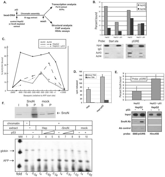
We purified the oncoprotein SnoN and found that it functions as a corepressor of the tumor suppressor p53 in the regulation of the hepatic alpha-fetoprotein (AFP) tumor marker gene. p53 promotes SnoN and histone deacetylase interaction at an overlapping Smad binding, p53 regulatory element (SBE/p53RE) in AFP. Comparison of wild-type and p53-null mouse liver tissue by using chromatin immunoprecipitation (ChIP) reveals that the absence of p53 protein correlates with the disappearance of SnoN at the SBE/p53RE and loss of AFP developmental repression. Treatment of AFP-expressing hepatoma cells with transforming growth factor-beta1 (TGF-beta1) induced SnoN transcription and Smad2 activation, concomitant with AFP repression. ChIP assays show that TGF-beta1 stimulates p53, Smad4, P-Smad2 binding, and histone H3K9 deacetylation and methylation, at the SBE/p53RE. Depletion, by small interfering RNA, of SnoN and/or p53 in hepatoma cells disrupted repression of AFP transcription. These findings support a model of cooperativity between p53 and TGF-beta effectors in chromatin modification and transcription repression of an oncodevelopmental tumor marker gene.


Figures

FIG. 1.
FIG. 1.
A TGF-β effector protein is purified from hepatic cells in the presence of the p53 protein. (A) Purification of SnoN protein. Solid-phase DNA oligomers, either biotinylated, 31-bp p53RE single-copy (lanes 3 to 6), ligated p53RE multimers (lanes 7 to 10), or single-copy, −1007 control (lanes 11 to 14) sequences, were incubated with 100 μg of total protein of adult mouse liver nuclear extract (ML, input extract; lane 2), in the presence of recombinant p53 (p53 alone; lane 1, recombinant protein stabilized by addition of BSA). Following binding, bead-DNA-protein complexes were washed, eluted, and then separated by denaturing gel electrophoresis alongside extract proteins not binding to DNA (unbound, lanes 3, 7, and 11). The gel was silver stained; eluted p53 (dagger, lanes 6 and 9) and a protein enriched in the presence of p53 (asterisk, lanes 5 and 9) were analyzed by MALDI mass spectrometry. (B) In vivo ChIP analysis of wild-type and p53-null mouse liver tissue. PCR analysis of antibody-precipitated genomic fragments was conducted with primers amplifying the region of the SBE/p53RE. Sonicated fragments of formaldehyde-cross-linked chromatin from 2-month-old wild-type (WT) and p53-null mouse liver tissue were antibody precipitated with tetra-acetylated histone H4 antibody (AcH4), Smad4, p53 (Ab-1), SnoN, P-Smad2, or control IgG. (C) Quantifying in vivo ChIP of Smad4 and P-Smad2 binding to the SBE/p53RE and distal genomic sites in WT and p53-null mouse liver tissue. Chromatin lysates were serially diluted (Input) and PCR amplified as described above to determine a linear range of amplification for all lysates. Antibody-precipitated fragments of DNA bound to Smad4 and SnoN were quantified by comparison to this range of values (% Bound). (D) Semiquantitative RT-PCR analysis of AFP RNA. RNA was isolated from 1- and 2-month-old WT and p53-null mouse liver tissue and analyzed in parallel for determination of GAPDH control RNA levels and AFP RNA levels.
FIG. 2.
FIG. 2.
SnoN, but not Ski, mediates repression of AFP expression. (A) SnoN-mediated transcription repression is compromised by disruption of p53 DNA binding. (Top) Hepa 1-6 hepatoma cells were transfected with AFP/lacZ (wild-type sequence for p53 binding) or DelA/lacZ (mutated for p53 binding) reporter constructs (500 ng/plate) along with the indicated expression vectors (p53 and SnoN; 500 ng/plate [each]). Each plate was also cotransfected with the pGL2 luciferase vector (50 ng/plate) to standardize and control for transfection efficiency. Expression levels relative to baseline are indicated for AFP/lacZ (gray) and DelA/lacZ (black). Cotransfected proteins exhibited equivalent levels of protein expression by Western blot analysis (data not shown). (Bottom) The p53/HNF-3 (FoxA) regulatory element centered at −850 in the AFP promoter (42) overlaps with consensus SBEs (89). The DelA/lacZ binding element has a 10-bp deletion within the 5′ p53 half site (shaded) and point mutations in the 3′ half site. (B) Ski activates AFP expression in the presence of p53. Identical transient transfection analyses were performed with the AFP/lacZ reporter and coexpressed p53, SnoN, and c-Ski constructs, as indicated. Expressed Ski protein repressed AFP only slightly and activated expression in the presence of p53. β-gal, β-galactosidase; luc, luciferase.
FIG. 3.
FIG. 3.
TGF-β represses endogenous AFP levels in hepatoma cells. (A) TGF-β and p53 repress AFP. Serum-starved Hepa 1-6 cells were treated with increasing concentrations of TGF-β1 (2 to 20 ng/ml of medium, lanes 3 to 7), vehicle (lanes 8 to 12), or no additions (lanes 1 and 2) and harvested at 0 h (lane 1) or 24 h (lanes 2 to 12) posttreatment. Whole-cell lysates were prepared from these cells, and immunoblot analyses were performed to analyze the effects of TGF-β1 treatment on AFP, p53, Smad4, and P-Smad2 protein levels. (B) AFP is a downstream physiological target of TGF-β. Hepa 1-6 cells were treated with 2 ng of TGF-β1/ml or vehicle control and harvested at the indicated time points posttreatment (0 to 56 h). Total RNA was harvested, and Northern blot analysis was performed to examine the levels of AFP and SnoN mRNA. The graph represents AFP (solid line) and SnoN (broken line) RNA levels in cells treated with ligand relative to those that received only the vehicle, each normalized to an actin RNA loading control. (C) TGF-β induces SnoN protein degradation, Smad2 protein phosphorylation, and a decrease in AFP. Hepa 1-6 cell lysates were treated with 4 ng of TGF-β1 ligand/ml or vehicle (V) only (as in panel B) over a 24-h interval and harvested at the indicated times for preparation of nuclear and cytoplasmic extracts. Immunoblot analysis of nuclear extracts was performed to determine SnoN, P-Smad2, p53, and actin protein levels. AFP levels were determined in cytoplasmic extracts of the same cells.
FIG. 4.
FIG. 4.
Downstream effectors of TGF-β and p53 are recruited to SBE/p53RE in vivo and effect chromatin modifications in response to TGF-β treatment. (A) Chromatin immunoprecipitation of proteins binding to the AFP SBE/p53RE. ChIP assays of SBE/p53RE were performed with Hepa 1-6 cells treated with a 2-ng/ml concentration of TGF-β1 (black bars) or vehicle control (gray bars) for 24 h. Immunoprecipitation of formaldehyde-cross-linked lysate was performed using 1.5 μg of antibody specific for p53, 3 μg of SnoN, 5 μg of Smad4, and 5 μl of P-Smad2 antibodies followed by PCR using oligonucleotides specific for the SBE/p53RE. As negative controls, lysis buffer alone was added to the immunoprecipitation or lysate was incubated with 5 μg of anti-IgG (see supplementary Fig. S2). PCRs using oligonucleotides specific for the AFP start site were also performed as an additional test for antibody specificity. Graphs indicate the average values of percent input bound calculated from multiple experiments and the standard error of the mean. n-fold changes of ligand versus vehicle control are shown below each data set. (B) Deacetylation and concomitant methylation of histone H3 lysine 9 is targeted at SBE/p53RE in response to TGF-β signaling. ChIP assays were performed as described above, except that 5 μg of anti-AcH4, 5 μg of anti-AcH3K9, and 5 μl of anti-diMetH3K9 antibodies were used for ChIP of formaldehyde-cross-linked chromatin lysates. Graphs indicate the average values of percent input bound calculated from multiple experiments and the standard errors of the means. n-fold changes are shown below each data set.
FIG. 5.
FIG. 5.
Both SnoN and p53 are essential for TGF-β-induced repression of AFP transcription. (A) Depletion of SnoN and p53 by siRNA methodology. Double-stranded oligomers homologous to specific sites within the coding regions of SnoN and p53, respectively, were cloned into U6 promoter-containing vectors and introduced by transfection of Hepa 1-6 cells. Three targets sequences for each gene were tested, each with various degrees of success in knockdown of protein expression. Left panel: transfection was performed without short, hairpin RNA-expressing constructs (shRNA; mock), in duplicate (a and b) with target 1 and separately with targets 2 and 3. Immunoblotting for SnoN revealed that targets 1 and 3 were most effective in knockdown of SnoN expression. Target 2 oligonucleotides contained sequence mismatches for the SnoN target region. Actin was analyzed as a control for nonspecific effects on total protein. Right panel: analysis of p53 knockdown was conducted on lysates of Hepa 1-6 cells treated with actinomycin D to activate p53 protein as previously described (42). Transfections of shRNA constructs were performed as described above. Lysate of actinomycin D-treated cells that were not transfected are analyzed in the lane labeled as “no siRNA. ” (B) Ligand-mediated repression of AFP transcription requires p53 and SnoN. Plasmid constructs capable of inducing siRNA for SnoN or p53 (target 1 constructs for each) were introduced separately or together into Hepa 1-6 cells by transient transfection. A vector specific for siRNA-mediated knockdown of firefly luciferase was used as a nonspecific, target control for RNA depletion. The “no siRNA” cells were not transfected. RNA was isolated from Hepa 1-6 cells, all of which were treated with TGFβ-1 ligand (2 ng/μl) or vehicle alone for 24 h, following a 24-h recovery period after transfection. Levels of AFP RNA were determined by Northern blot analysis and comparison to a cyclophilin RNA loading control. Left panel: a graph summarizing the results of four siRNA experiments; RNA levels were determined by Northern blot as described above. Error bars indicate standard deviation. Right panel: representative Northern blot of AFP and cyclophilin RNA isolated from vehicle only (V) or TGF-β1 ligand-treated (L) cells, which received siRNA constructs as indicated. (C) Immunoblot analysis of siRNA-treated nuclear extracts. SiRNA and ligand or vehicle-only conditions were exactly as shown in panel B and as described above. Actin protein analysis serves as a loading control and a control for nonspecific effects of siRNA treatment.
FIG. 6.
FIG. 6.
p53 targets histone deacetylation to the SBE/p53RE and mediates SnoN-dependent repression of AFP transcription. (A) Diagram of in vitro chromatin assembly and analysis. AFP bead DNA is preincubated with protein extracts and/or recombinant p53 protein prior to chromatin assembly over a 1-h incubation in X. laevis (Xl) egg extract. Assembled chromatin bead DNA is then washed in an isotonic buffer prior to functional assay by transcription in HeLa extract or structural analysis by ChIP and HDAC assays. (B and C) p53 triggers deacetylation of local and distal chromatin. ChIP assays (two to three independent assays for each data point) were performed in vitro on AFP/lacZ DNA, which was chromatin assembled in the presence of HepG2 whole-cell extract or HepG2 plus 500 ng of activated p53. DNA was purified from protein-chromatin immunoprecipitates and analyzed for percent SBE/p53RE and transcription start site DNA bound to acetylated histone H3 (AcH3) or histone H4 (AcH4) by Southern blotting with labeled (B) SBE/p53RE or start site (+4 to +26) oligonucleotides or (C) across the AFP templates with oligonucleotides specific for +10/+32, −140/−118, −327/−305, −520/−398, −800/−778, −2637/−2615, and +3339/+3361. Representative data are shown below the summary graph (B). (D) p53-dependent HDAC activity at SBE/p53RE. Immobilized SBE/p53RE DNA oligomers were incubated in 100 μg of HepG2 extract, either tryptic soy agar treated (10 μM; black bars) or untreated (gray bars), in the presence or absence of recombinant, activated p53. Protein complexes bound to the oligomers were washed and incubated with in vivo-labeled, 3H-acetylated histones, which were isolated and purified. HDAC enzymatic activity was measured by scintillation counting of released [3H]acetyl groups. The graphrepresents average values from multiple experiments and standard deviations of the means. (E) p53-dependent SnoN association at the SBE/p53RE. A series of ChIP studies (≥3 for each data point) were performed as described above. Values for percentage of SnoN bound relative to input at the SBE/p53RE in the presence and absence of recombinant p53 were normalized to antibody (Ab) control reactions. Probes used for Southern blots of precipitated DNA were labeled SBE/p53RE oligonucleotides or Hinc450 oligonucleotides (+3333 to +3356), which will detect nonspecific binding to AFP/lacZ DNA. (F) SnoN is required for p53-mediated repression of in vitro AFP chromatin transcription. HepG2 whole-cell extract was immunodepleted for SnoN protein (upper panel) and then analyzed by in vitro chromatin transcription (lower panel). Upper panel: 40 μg of protein of starting HepG2 extract, input (I), equal levels of protein from depleted extract, supernatant (S) and pellet (P) derived from boiled antibody-coupled beads used for immunodepletion. Lower panel: immobilized AFP DNA templates were incubated in (i) control (lanes 3 to 5; approximately 200 μg of total protein), (ii) immunodepleted (lanes 6 to 8; approximately 200 μg of total protein), or (iii) mock-depleted (lanes 9 and 10; approximately 200 μg of total protein) extracts or (iv) buffer (lanes 1 and 2) in the presence of recombinant p53 (500 ng, lanes 4 and 7; 1 μg, lanes 5, 8, and 10) or p53 dialysis buffer (lanes 1 to 3, 6 and 9) prior to chromatin assembly. Chromatin-assembled DNA templates were washed in nuclear extract buffer and in vitro transcribed in HeLa nuclear extract. Chick β-globin (50 ng) DNA was added during the transcription period as an RNA recovery control (57). AFP and Globin primer extension products are indicated. n-fold changes of transcribed AFP in vitro RNA signals are shown below each data point.

Similar articles

Cited by

References

    1. Akiyoshi, S., H. Inoue, J. Hanai, K. Kusanagi, N. Nemoto, K. Miyazono, and M. Kawabata. 1999. c-Ski acts as a transcriptional co-repressor in transforming growth factor-beta signaling through interaction with smads. J. Biol. Chem. 274:35269-35277. - PubMed
    1. Andrisani, O. M., and S. Barnabas. 1999. The transcriptional function of the hepatitis B virus X protein and its role in hepatocarcinogenesis. Int. J. Oncol. 15:373-379. - PubMed
    1. Ashcroft, M., Y. Taya, and K. H. Vousden. 2000. Stress signals utilize multiple pathways to stabilize p53. Mol. Cell. Biol. 20:3224-3233. - PMC - PubMed
    1. Attisano, L. 2000. Smads as transcriptional co-modulators. Curr. Opin. Cell Biol. 12:235-243. - PubMed
    1. Bargonetti, J., and J. J. Manfredi. 2002. Multiple roles of the tumor suppressor p53. Curr. Opin. Oncol. 14:86-91. - PubMed

Publication types

MeSH terms