. 2005;33(1):43-55.
doi: 10.1093/nar/gki143.

New insights into DNA triplexes: residual twist and radial difference as measures of base triplet non-isomorphism and their implication to sequence-dependent non-uniform DNA triplex

Affiliations

New insights into DNA triplexes: residual twist and radial difference as measures of base triplet non-isomorphism and their implication to sequence-dependent non-uniform DNA triplex

R Thenmalarchelvi et al. Nucleic Acids Res. 2005.

Abstract

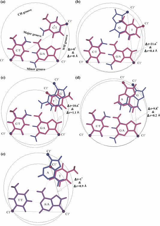
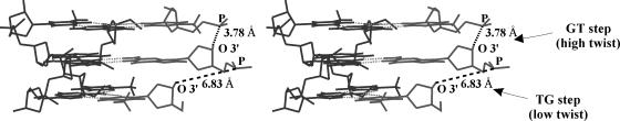
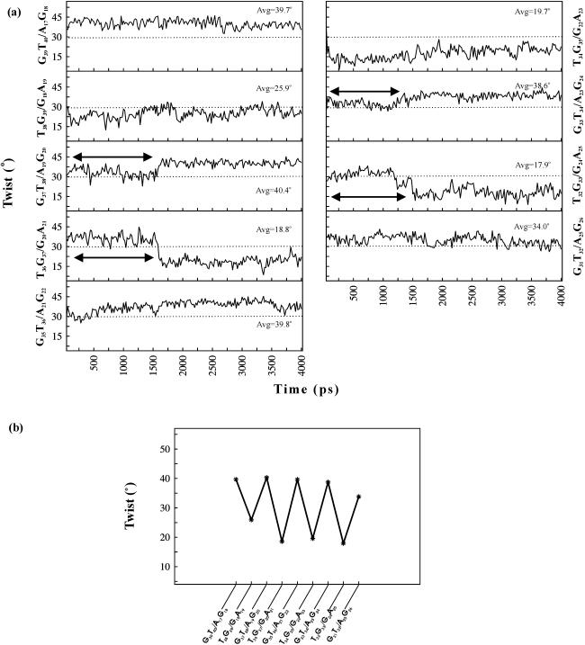
DNA triplexes are formed by both isomorphic (structurally alike) and non-isomorphic (structurally dissimilar) base triplets. It is espoused here that (i) the base triplet non-isomorphism may be articulated in structural terms by a residual twist (Delta(t) degrees), the angle formed by line joining the C1'...C1' atoms of the adjacent Hoogsteen or reverse Hoogsteen (RH) base pairs and the difference in base triplet radius (Delta(r) A), and (ii) their influence on DNA triplex is largely mechanistic, leading to the prediction of a high (t + Delta(t))degrees and low (t - Deltat)degrees twist at the successive steps of Hoogsteen or RH duplex of a parallel or antiparallel triplex. Efficacy of this concept is corroborated by molecular dynamics (MD) simulation of an antiparallel DNA triplex comprising alternating non-isomorphic G*GC and T*AT triplets. Conformational changes necessitated by base triplet non-isomorphism are found to induce an alternating (i) high anti and anti glycosyl and (ii) BII and an unusual BIII conformation resulting in a zigzag backbone for the RH strand. Thus, base triplet non-isomorphism causes DNA triplexes into exhibiting sequence-dependent non-uniform conformation. Such structural variations may be relevant in deciphering the specificity of interaction with DNA triplex binding proteins. Seemingly then, residual twist (Delta(t) degrees) and radial difference (Deltar A) suffice as indices to define and monitor the effect of base triplet non-isomorphism in nucleic acid triplexes.


Figures

Figure 1
Figure 1
Definition of intrinsic residual Hoogsteen twist and residual reverse Hoogsteen (Δt°) twist. Superposition of (a) isomorphic T*AT (maroon) and C+*GC (blue) (parallel orientation) and non-isomorphic base triplets: (b) G*GC (blue) and T*AT (maroon) (parallel orientation) (c) G*GC (blue) and T*AT (maroon) (antiparallel orientation) (d) G*GC (blue) and A*AT (maroon) (antiparallel orientation) and (e) A*AT (blue) and T*AT (maroon) (antiparallel orientation). Dotted lines represent the hydrogen bonds. Filled circles represent the C1′ atoms. Coincidence of C1′ atoms and the lines joining C1′…C1′ atoms of WC and Hoogsteen base pairs is seen only in (a). Non-coincidence of all the C1′ atoms leads to the formation of an angle between the lines joining C1′…C1′ atoms of the Hoogsteen and reverse Hoogsteen base pairs (b–d). This is tantamount to a pre-existing helical twist and is referred to as residual Hoogsteen twist or residual reverse Hoogsteen twist, and is denoted by ‘Δt°’. Difference in triplet radii (Δr Å) reflects yet another factor contributing to base triplet non-isomorphism (c and e), besides ‘Δt°’.
Scheme 1
Scheme 1
A 14mer antiparallel DNA triplex with alternating non-isomorphic G*GC and T*AT triplets. Base sequence is numbered to facilitate discussion. RH hydrogen bond pairs are represented by ‘*’. Here onwards, the pyrimidine and purine strands of the WC duplex is referred to as Crick and Watson strand, respectively, and the third strand is referred to as the reverse Hoogsteen (RH) strand.
Figure 2
Figure 2
Stereo diagram showing the disjointed nature of the reverse Hoogsteen (RH) strand (grey) caused by Δt = 10.6° and Δr Å in an antiparallel DNA triplex (t = 30° and h = 3.26 Å) comprising alternating G*GC and T*AT triplets. Gaps of nearly one nucleotide length at TG step may be seen. WC duplex is coloured black.
Figure 3
Figure 3
(a) Variation of helical twist angles at the alternating steps of the central 10mer of the RH d(GT)7*d(AG)7 duplex over 4 ns dynamics. Triplex structure at every 20 ps interval is considered for calculation of twists. Note the alternating high and low twist at alternating steps. Dotted lines correspond to the assigned twist angle of 30°. Double-headed arrows indicate unusual variations seen at different steps. Average values of twist calculated over the last 2.4 ns are also given. (b) Variations of helical twist angles at different steps of the average structure of the central 10mer RH duplex. Alternating high and low twist angles at GT/AG and TG/GA steps result in a saw–tooth-like pattern. Base sequences corresponding to different steps of the RH duplex are shown for clarity. However, base sequence along the RH strand alone is referred in the text.
Figure 4
Figure 4
Variation of conformational angles ɛ(C3′–O3′), ζ(O3′–P) and χ(C1′–N9/N1) and P (phase angle of pseudo rotation) in the central 10mer of the RH strand over 4 ns dynamics. Alternating high anti (G) and anti (T) glycosyl conformation along with alternating BII (GT step) and BIII (TG step) conformation may be seen.
Figure 5
Figure 5
Variation of N3(T36)…N7(A21) and O2(T36)…N6(A21) RH hydrogen bond distances over 4 ns simulation. Note the large fluctuation in N3(T36)…N7(A21) hydrogen bond distance during the first 400 ps, and in O2(T36)…N6(A21) hydrogen bond distance during the first 1600 ps. These are correlated with water-mediated interactions shown in Figure 6 (see also text). Hydrogen bond distances at every 1 ps interval are calculated.
Figure 6
Figure 6
Interaction of water with RH T36…A21 pair during the dynamics. The spikes in Figure 5 correspond to a situation when water comes exactly between A21 and T36 around 400 ps. Water molecules are represented by ball and stick.
Figure 7
Figure 7
(a) Stereo plot of the antiparallel DNA triplex comprising alternating G*GC and T*AT base triplets corresponding to the last 2.4 ns of the central 10mer of the average structure. Smooth curve linking the phosphates of the WC duplex and the zig-zag nature of the RH strand (green) may be readily seen. (b) RMS deviation (r.m.s.d.) of the central 10mer triplex structures from the average structure. Triplex structures at every 1 ps interval over 4 ns dynamics are taken for the calculation. Note the stabilization of the RMS deviation after 3 ns.
Figure 8
Figure 8
Stacking interactions at the G35T36 step (G35*G22C7/T36*A21T8) and T36G37 step (T36*A21T8/G37*G20C9) of the average structure. Hydrogen bonds are indicated by dotted lines. Figures are drawn using 3DNA (32).
Figure 9
Figure 9
Schematic diagram showing the water network around the tetrameric triplex T34*A23T6–G35*G22C7–T36*A21T8–G37*G20C9. Donor…acceptor distances within 3.5 Å are shown by dotted lines. Filled circles (grey) indicate water molecules.
Figure 10
Figure 10
Stereo diagram showing the BI (a and b), BII (c and d) and BIII (e and f) conformation that occurs at the low twist TG step of the RH strand during the dynamics.

Similar articles

Cited by

References

    1. Soyfer V.N., Potaman V.N. Triple-Helical Nucleic Acids. New York: Springer-Verlag; 1996.
    1. Gowers D.M., Fox K.R. Towards mixed sequence recognition by triple helix formation. Nucleic Acids Res. 1999;27:1569–1577. - PMC - PubMed
    1. Praseuth D., Guieysse A.L., Helene C. Triple helix formation and the antigene strategy for sequence-specific control of gene expression. Biochim. Biophys. Acta. 1999;1489:181–206. - PubMed
    1. Wang E., Feigon J. Structures of nucleic acid triplexes. In: Neidle S., editor. Oxford Handbook of Nucleic Acid Structure. New York: Oxford University Press; 1999. pp. 355–388.
    1. Vasquez K.M., Glazer P.M. Triplex-forming oligonucleotides: principles and applications. Q. Rev. Biophys. 2002;35:89–107. - PubMed

Publication types