. 2005 Mar;128(Pt 3):516-27.
doi: 10.1093/brain/awh390. Epub 2005 Jan 19.

Early and late HHV-6 gene transcripts in multiple sclerosis lesions and normal appearing white matter

Affiliations

Early and late HHV-6 gene transcripts in multiple sclerosis lesions and normal appearing white matter

Margaret L Opsahl et al. Brain. 2005 Mar.

Abstract

Multiple sclerosis is an inflammatory demyelinating disease of the CNS, the aetiology of which is believed to have both genetic and environmental components. We have investigated one of the candidate viruses for the environmental component of multiple sclerosis, the neurotropic human herpesvirus 6 (HHV-6). Utilizing fluorescent in situ hybridization (FISH) techniques, we have examined human post-mortem tissues for the presence of immediate early and late viral gene expression in multiple sclerosis patient normal appearing white matter (NAWM), lesional tissue and normal control brain samples. HHV-6 gene transcription was detected in all tissue samples and was restricted to oligodendrocytes, as determined by double mRNA FISH analysis. Quantitative analysis of viral mRNA expression indicated that both NAWM and lesional multiple sclerosis samples exhibited significantly higher levels of HHV-6 expression compared with the normal control samples. Lesional samples exhibited the highest levels of viral gene expression, with NAWM exhibiting an intermediate level between lesional and control tissues. Immunofluorescence against early and late HHV-6 proteins verified active translation of HHV-6 viral mRNA in oligodendrocytes. Southern blot analysis of nested polymerase chain reactions using extracted genomic DNA and cDNA confirmed the presence of the HHV-6 genome in all individuals, with the active expression profile mirroring the FISH results. The frequent high level of HHV-6 infection in multiple sclerosis samples suggests a possible role in pathogenesis.


Figures
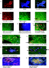
Fig. 1
Fig. 1
Both immediate early and late viral gene expression is seen in all samples and locates to the cytoplasm of oligodendrocytes, indicating an active infection. The number of cells that are positive for HHV-6 mRNA varies but cells tend to be seen in clusters. (A) Representative FISH of claudin-11 mRNA (rhodamine red), ISH for myelin oligodendrocyte basic protein (MOBP) using AMCA (blue)-conjugated secondary antibody with the two colour images merged to demonstrate co-localization of both signals indicating that the mRNA probe correctly identifies oligodendrocytes (magnification ×400). The image is taken from normal control tissue. (B) Representative double FISH for claudin-11 mRNA (rhodamine red), HHV-6 mRNA (fluorescein) and the two colour merged image demonstrating co-localization of both signals to oligodendrocyte cells (orange). The circle and arrow indicate two representative cells showing first claudin-11 mRNA, then HHV-6 mRNA and finally the two merged images (magnification ×200). The image is taken from multiple sclerosis NAWM tissue. (C) Normal control tissue probed with sense probes for HHV-6 (fluorescein) and claudin-11 (rhodamine red). All that can be seen are the DAPI-counterstained nuclei (magnification ×200). (D) Mouse brain section probed for HHV-6 mRNA (fluorescein), claudin-11 mRNA (rhodamine red) and MOBP protein (AMCA secondary). Only MOBP signal is present. The circle and arrow indicate a representative oligodendrocyte (magnification ×200). (E) Representative ISH for GFAP (fluorescein secondary) and claudin-11 mRNA FISH (rhodamine red). The signals do not co-localize, the circle and arrows highlighting examples of both (magnification ×200). The image is taken from normal control tissue. (F) Representative double FISH for claudin-11 and HHV-6 immediate early mRNA with both signals co-localizing (orange). The circle and arrow indicate two representative cells where the two mRNA signals co-localize (magnification ×200). (G) Representative double FISH for claudin-11 and HHV-6 late mRNA with both signals co-localizing (orange). The circle and arrow indicate two representative cells where the two mRNA signals co-localize (magnification ×200). (H) Representative double ISH for MOBP (AMCA secondary) and HHV-6 immediate early (rhodamine secondary) proteins exhibiting co-localization of the signal to oligodendrocytes (purple) confirming mRNA FISH results (magnification ×400). (I) Representative double ISH for MOBP (AMCA secondary) and HHV-6 late (rhodamine secondary) proteins exhibiting co-localization of signal to oligodendrocytes (purple) confirming mRNA FISH results. The circle and arrow indicate two representative cells where the two protein signals co-localize (magnification ×200). Images FI are from from multiple sclerosis lesion tissue. (J) Representative ISH for HHV-6A protein (rhodamine secondary) and HHV-6 mRNA FISH (fluorescein) (magnification ×400). The image is taken from multiple sclerosis NAWM. (K) Representative ISH for HHV-6B protein (rhodamine secondary) and HHV-6 mRNA FISH (fluorescein) (magnification ×400). The image is taken from multiple sclerosis NAWM.
Fig. 2
Fig. 2
Graphical representation of the data presented in Table 1.
Fig. 3
Fig. 3
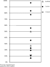
Scatterplot representing the data presented in Table 2. Each point represents an individual sample. The control sample value is represented by a broad line, lesional samples are filled diamonds and NAWM are open triangles. As two of the control samples had undetectable HHV-6 mRNA, only the third sample appears on this graph representing what has to be considered a normal level of HHV-6 infection in oligodendrocytes.
Fig. 4
Fig. 4
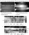
RT–PCR of snap-frozen samples for (A) the endogenous housekeeping gene β-microglobulin (250 bases) and (B) the oligodendrocyte-specific gene claudin-11 (500 bases). Note that not all samples are positive for cDNA, even for the housekeeping gene, with claudin-11 less abundant and more variable. Both are 2% agarose gels with a 1 kb ladder in increments of 100 bases. N = NAWM; L = lesion. As both fixed frozen and snap-frozen samples were not available for all multiple sclerosis patients, some of the PCR samples will not be seen with FISH data and vice versa. Southern blot of genomic DNA (C) and cDNA (D) for the HHV-6 IE gene using nested PCR demonstrates the presence of the viral genome in all samples (highlighted box in C) and transcription in most sample sets (arrows highlight the strongest bands), complementing mRNA FISH and protein data. As nested PCR was utilized, three bands are seen in the positive control lanes representing the three possible primer combinations. The main middle band is predominant in the genomic DNA samples (C), whilst examples of all three are evident in the cDNA samples (D).

Similar articles

Cited by

References

    1. Ablashi DV, Balanchandran N, Josephs SF, Hung CL, Krueger GR, Kramarsky B, et al. Genomic polymorphism, growth properties and immunologic variations in human herpesvirus-6 isolates. Virology 1991; 184: 545–52. - PubMed
    1. Ablashi DV, Lapps W, Kaplan M, Whitman JE, Richert JR, Pearson GR. Human herpesvirus-6 (HHV-6) infection in multiple sclerosis: a preliminary report. Mult Scler 1998; 4: 490–6. - PubMed
    1. Ablashi DV, Eastman HB, Owen CB, Roman MM, Friedman J, Zabriskie JB, et al. Frequent HHV-6 reactivation in multiple sclerosis (MS) and chronic fatigue syndrome (CFS) patients. J Clin Virol 2000; 16: 179–91. - PubMed
    1. Akhyani N, Berti R, Brennan MB, Soldan SS, Eaton JM, McFarland, et al. Tissue distribution and variant characterization of human herpesvirus (HHV)-6: increased prevalence of HHV-6A in patients with multiple sclerosis. J Infect Dis 2000; 182: 1321–25. - PubMed
    1. Albright AV, Lavi E, Black JB, Goldberg S, O'Connor MJ, Gonzalez-Scarano F. The effect of human herpesvirus-6 (HHV-6) on cultured human neural cells: oligodendrocytes and microglia. J Neurovirol 1998; 4: 486–94. - PubMed

Publication types

MeSH terms