

doi: 10.1073/pnas.0409217102.
Epub 2005 Jan 19.
Receptor editing in peripheral B cell tolerance
Affiliations
- PMID: 15659547
- PMCID: PMC547880
- DOI: 10.1073/pnas.0409217102
Item in Clipboard
Receptor editing in peripheral B cell tolerance
Proc Natl Acad Sci U S A.
.
Abstract
Receptor editing or secondary Ig gene rearrangement occurs in immature, autoreactive B cells to maintain self-tolerance. Here we show that nonspontaneously autoimmune mice immunized with a peptide mimetope of DNA develop peptide- and DNA-reactive antibodies. Antigen-specific B cells display a follicular B cell phenotype. As these cells move into the memory compartment, many express RAG protein and acquire expression of both kappa and lambda light chains. Thus, this study provides evidence for receptor editing occurring in a mature, antigen-activated B cell population. Because the receptor editing observed here occurred in an autoreactive response to antigen, it may function to maintain peripheral tolerance.
Figures
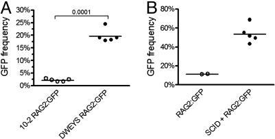
RAG expression in autoreactive, mature B cells. (A) GFP expression in B220+HSAlowtetramer+ B cells from DWEYS (filled circles) and 10-2 (open circles) immunized RAG2:GFP Tg mice. (B) GFP expression in B220+HSAlowtetramer+ B cells after adoptive transfer and challenge.
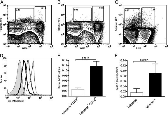
Differentiation and maturity of tetramer-reactive splenocytes. (A–C) Representative tetramer staining after secondary challenge in the spleen (A) and inhibition of splenic staining by 30 mM nonfluorescent tetramer (B) and bone marrow (C). (D) Histogram of intracellular IgG staining in tetramer populations. Thick line, tetramer+B220low; thin line, tetramer+B220hi; dotted line, tetramer–B220hi; shaded line, tetramer–B220–. (E) Expression of AID in tetramer populations as determined by real-time PCR. (F) Expression of Bcl-6 in tetramer populations as determined by real-time PCR. Data in A–D are representative of 10 mice. Real-time PCR in E and F is representative of three independent experiments.
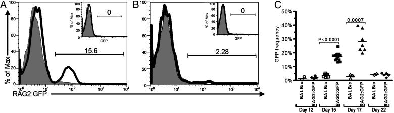
RAG expression in tetramer-reactive B cells. (A and B) Representative histogram of GFP expression on day 15 in B220+HSAlow DWEYS+ (A) and 10-2+ (B) B cells from RAG2:GFP Tg mice (solid line) compared to B220+HSAlowtetramer+ B cells in wild-type mice (shaded line). GFP expression is not seen in B220+HSAlowtetramer– B cells in RAG2:GFP Tg mice (Insets). (C) Time course of GFP expression in DWEYS-immunized RAG2:GFP Tg mice versus wild-type mice.
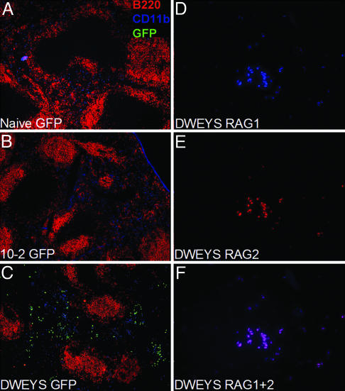
Histological analysis of RAG2:GFP and BALB/c mice. (A–C) RAG2:GFP+/– spleen sections stained with anti-B220 (red), anti-CD11b (blue), and anti-GFP (green) in naive (A), 10-2-immunized (B), and DWEYS-immunized (C) mice. Images were obtained by using the ×10 objective. (D–F) BALB/c spleen sections from DWEYS-immunized mice stained with anti-RAG1 (D) or anti-RAG2 (E), and superimposed (F). Images were obtained by using the ×40 objective.
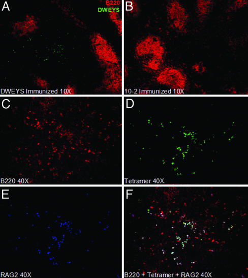
Histological staining with fluorescent tetramers. (A and B) BALB/c spleen sections from 10-2 (A) or DWEYS (B) immunized mice stained with anti-B220 (red) and DWEYS-A488 (green). Images were obtained by using the ×10 objective. (C–F) BALB/c spleen sections from DWEYS immunized mice stained with anti-B220 (C, red), DWEYS-A488 (D, green), anti-RAG2 (E, blue), and superimposed (F). Images were obtained by using the ×40 objective.
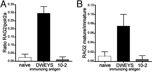
Real-time PCR analysis of RAG2 expression and analysis of receptor editing. (A) Real-time PCR analysis of RAG2 expression versus RNA polymerase 2 (housekeeping gene) in mature B cells (B220+HSAlowAA4.1low) from naive, DWEYS-, or 10-2-immunized mice. (B) Ratio of RAG2 expression in mature versus immature (B220+HSAhiAA4.1hi) B cells in naive, DWEYS-, or 10-2-immunized mice.
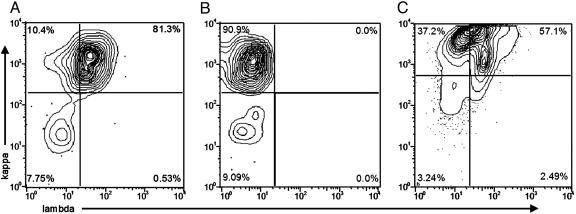
Analysis of light chain expression. (A) Bivariate plots depicting λ and κ staining of DWEYS tetramer-gated cells. (B) Bivariate plots depicting κ and λ staining of 10-2 tetramer-gated cells. (C) Bivariate plots depicting λ and κ staining of DWEYS tetramer gated cells from SCID recipients of BALB/c splenocytes.
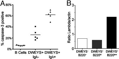
Expression of activated caspase-3 and λ light chain. (A) Active caspase-3 expression in B220+ (circles), DWEYS+λ– (squares), and DWEYS+λ+ B cells (triangles). (B) Densitometric quantitation of λ light chain/actin RNA as determined by semiquantitative RT-PCR on the indicated sorted populations.
References
-
- Benschop, R. J., Aviszus, K., Zhang, X., Manser, T., Cambier, J. C. & Wysocki, L. J. (2001) Immunity 14, 33–43. - PubMed
-
- Hande, S., Notidis, E. & Manser, T. (1998) Immunity 8, 189–198. - PubMed
-
- Klinman, D. M., Shirai, A., Ishigatsubo, Y., Conover, J. & Steinberg, A. D. (1991) Arthritis Rheum. 34, 1404–1410. - PubMed
MeSH terms
Substances
LinkOut - more resources
Full Text Sources
Research Materials
