. 2005 Jan 19;33(2):478-85.
doi: 10.1093/nar/gki194. Print 2005.

Ribosomal protein L1 recognizes the same specific structural motif in its target sites on the autoregulatory mRNA and 23S rRNA

Affiliations

Ribosomal protein L1 recognizes the same specific structural motif in its target sites on the autoregulatory mRNA and 23S rRNA

Natalia Nevskaya et al. Nucleic Acids Res. .

Abstract

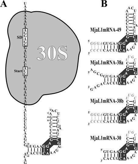
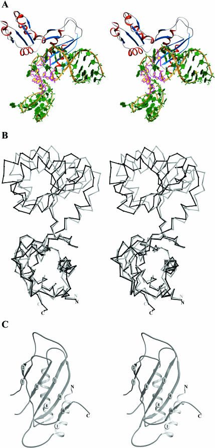
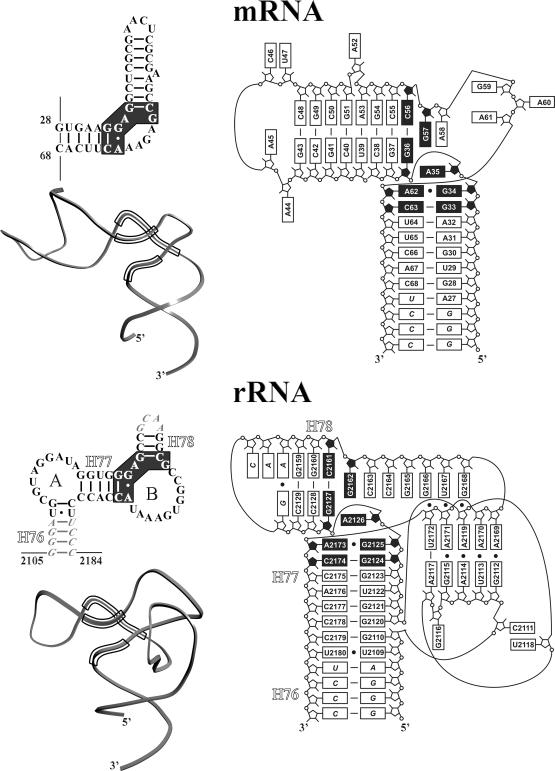
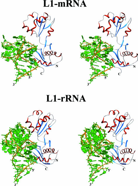
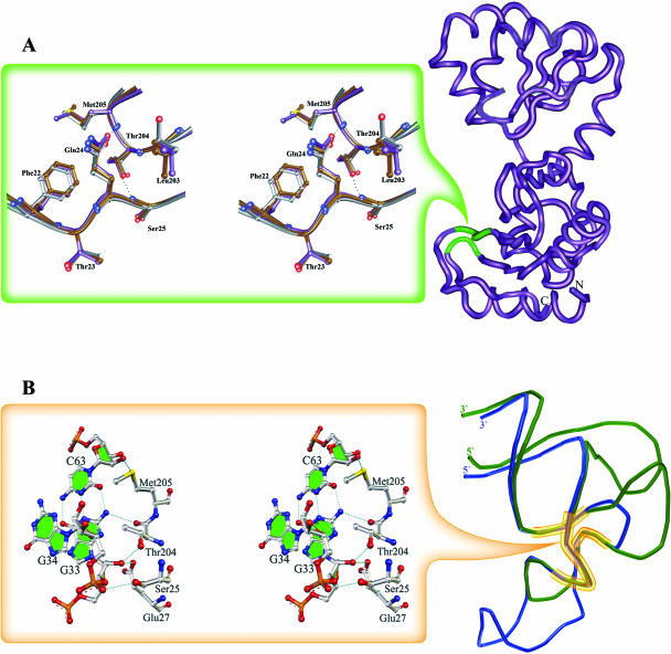
The RNA-binding ability of ribosomal protein L1 is of profound interest since the protein has a dual function as a ribosomal protein binding rRNA and as a translational repressor binding its mRNA. Here, we report the crystal structure of ribosomal protein L1 in complex with a specific fragment of its mRNA and compare it with the structure of L1 in complex with a specific fragment of 23S rRNA determined earlier. In both complexes, a strongly conserved RNA structural motif is involved in L1 binding through a conserved network of RNA-protein H-bonds inaccessible to the solvent. These interactions should be responsible for specific recognition between the protein and RNA. A large number of additional non-conserved RNA-protein H-bonds stabilizes both complexes. The added contribution of these non-conserved H-bonds makes the ribosomal complex much more stable than the regulatory one.


Figures

Figure 1
Figure 1
Secondary structure of the regulatory L1-binding site on the L1mRNA of M.jannaschii and derivatives thereof used in binding experiments and crystallization trials. (A) Localization of the L1-binding site and of the 30S ribosomal subunit as part of the translation initiation complex on the L1mRNA. Nucleotides different in the M.vannielii L1mRNA are shown with smaller sized letters. (B) Fragment MjaL1mRNA-49 comprising nucleotides +28 to +68 with four additional base pairs (italic) at the 3′ and the 5′ end. Fragment MjaL1mRNA-38a comprising nucleotides +24 to +38 and +54 to +72 is closed by the tetraloop UUGC (italic). Fragment MjaL1mRNA-38b is similar to MjaL1mRNA-38a, but the free 3′ and 5′ ends have been replaced by an extended helix as in MjaL1mRNA-49. Fragment MjaL1mRNA-30 comprising nucleotides +28 to +38 and +54 to +68 is closed by a tetraloop as in MjaL1mRNA-38. The cluster of nucleotides conserved in all 23S rRNA and mRNA binding sites is shown with black background.
Figure 2
Figure 2
(A) Stereo view of the L1–mRNA complex. The ribose-phosphate backbone is in gold, bases are in green, β-strands in blue and α-helices in red. Conserved nucleotides are shown in magenta and yellow. (B) Superposition of the structures of the isolated MjaL1 protein (gray) and MjaL1 from the present complex (black) with least squares minimization of differences in Cα atom coordinates of domain I. (C) Stereo view of the MjaL1 domain I.
Figure 3
Figure 3
Secondary structures, ribbon representation and schematic illustration of three-dimensional structures of the mRNA and rRNA fragments. Nucleotide replacements, which were introduced to the RNA fragments to facilitate the complex crystallization, are in italic. Conserved nucleotides are shown with black background.
Figure 4
Figure 4
Stereo ribbon representation of the L1–mRNA and L1–rRNA complexes in the same orientation.
Figure 5
Figure 5
Invariant regions in L1 proteins and in the rRNA and mRNA fragments. Conserved H-bonds are shown with dotted lines. (A) The location of the invariant structure on the surface of L1 proteins is shown in green on the MjaL1 model (right). Superposition of the corresponding regions in L1 proteins in isolated and RNA-bound forms is shown on the left. Isolated MjaL1 is in gray, MjaL1 complexed with mRNA in magenta and SacL1 complexed with rRNA in brown. (B) Superposition of the mRNA (blue) and rRNA (green) fragments; the conserved unique structure is outlined (right).

References

    1. Nikonov S., Nevskaya N., Eliseikina I., Fomenkova N., Nikulin A., Ossina N., Garber M., Jonsson B.-H, Briand C., Svensson A., Aevarsson A., Liljas A. Crystal structure of the RNA-binding ribosomal protein L1 from Thermus thermophilus. EMBO J. 1996;15:1350–1359. - PMC - PubMed
    1. Nevskaya N., Tishchenko S., Fedorov R., Al-Karadaghi S., Liljas A., Kraft A., Piendl W., Garber M., Nikonov S. Archaeal ribosomal protein L1: the structure provides new insights into RNA binding of the L1 protein family. Structure. 2000;8:363–371. - PubMed
    1. Nevskaya N., Tishchenko S., Paveliev M., Smolinskaya Y., Fedorov R., Piendl W., Nakamura Y., Toyoda T., Garber M., Nikonov S. Structure of ribosomal protein L1 from Methanococcus thermolithotrophicus. Functionally important structural invariants on the L1 surface. Acta Crystallogr. 2002;D58:1023–1029. - PubMed
    1. Zimmermann R.A. Interactions among protein and RNA components of the ribosome. In: Chambliss G., Craven G., Davies J., Davies K., Kahan L., Nomura M., editors. Ribosomes. Structure, Function and Genetics. Baltimore: University Park Press; 1980. pp. 135–169.
    1. Nikulin A., Eliseikina I., Tishchenko S., Nevskaya N., Davydova N., Platonova O., Piendl W., Selmer M., Liljas A., Drygin D., Zimmermann R., Garber M., Nikonov S. Structure of the L1 protuberance in the ribosome. Nature Struct. Biol. 2003;10:104–108. - PubMed

Publication types

MeSH terms

Associated data