. 2005 Jan 19;25(3):619-28.
doi: 10.1523/JNEUROSCI.3959-04.2005.

Glial cell line-derived neurotrophic factor mediates the desirable actions of the anti-addiction drug ibogaine against alcohol consumption

Affiliations

Glial cell line-derived neurotrophic factor mediates the desirable actions of the anti-addiction drug ibogaine against alcohol consumption

Dao-Yao He et al. J Neurosci. .

Abstract

Alcohol addiction manifests as uncontrolled drinking despite negative consequences. Few medications are available to treat the disorder. Anecdotal reports suggest that ibogaine, a natural alkaloid, reverses behaviors associated with addiction including alcoholism; however, because of side effects, ibogaine is not used clinically. In this study, we first characterized the actions of ibogaine on ethanol self-administration in rodents. Ibogaine decreased ethanol intake by rats in two-bottle choice and operant self-administration paradigms. Ibogaine also reduced operant self-administration of ethanol in a relapse model. Next, we identified a molecular mechanism that mediates the desirable activities of ibogaine on ethanol intake. Microinjection of ibogaine into the ventral tegmental area (VTA), but not the substantia nigra, reduced self-administration of ethanol, and systemic administration of ibogaine increased the expression of glial cell line-derived neurotrophic factor (GDNF) in a midbrain region that includes the VTA. In dopaminergic neuron-like SHSY5Y cells, ibogaine treatment upregulated the GDNF pathway as indicated by increases in phosphorylation of the GDNF receptor, Ret, and the downstream kinase, ERK1 (extracellular signal-regulated kinase 1). Finally, the ibogaine-mediated decrease in ethanol self-administration was mimicked by intra-VTA microinjection of GDNF and was reduced by intra-VTA delivery of anti-GDNF neutralizing antibodies. Together, these results suggest that GDNF in the VTA mediates the action of ibogaine on ethanol consumption. These findings highlight the importance of GDNF as a new target for drug development for alcoholism that may mimic the effect of ibogaine against alcohol consumption but avoid the negative side effects.


Figures
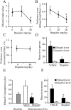
Figure 1.
Figure 1.
Ibogaine decreases ethanol consumption. A, Ibogaine decreased ethanol consumption expressed as mean ± SEM grams of ethanol per kilogram body weight during continuous access to both ethanol and water when intake was measured 24 hr after an acute injection (F(2,16) = 5.12; p < 0.02). *p < 0.05 compared with vehicle treatment (n = 9). B, Ibogaine treatment decreased ethanol preference expressed as mean milliliters of ethanol/(ml ethanol + ml water) ± SEM (F(2,16) = 7.83; p < 0.005). *p < 0.05 compared with vehicle treatment (n = 9). C, Ibogaine did not affect sucrose preference, expressed as mean milliliters of sucrose/(ml sucrose + ml water) ± SEM, measured 24 hr after injection (F(1,8) = 0.02; p = 0.88) (n = 9). D, Ibogaine attenuated operant ethanol self-administration in rats. Systemic ibogaine injected 3 hr before the session reduced responding for oral ethanol at the ethanol-paired lever but not the inactive lever (main effect of treatment: F(1,7) = 5.77, p < 0.05; main effect of lever: F(1,7) = 15.83, p < 0.006; treatment × lever interaction: F(1,7) = 5.78, p < 0.05]. Ethanol was delivered on an FR3 reinforcement schedule. Data are shown as mean ± SEM. *p < 0.05 compared with active lever responding after vehicle treatment (n = 8). E, Enhanced ethanol intake after a period of extinction was reduced by ibogaine injected 3 hr before the reinstatement test session (main effect of treatment: F(2,12) = 10.07, p < 0.03). Data are shown as mean ± SEM. *p < 0.02 compared with baseline responding before extinction; #p < 0.002 compared with vehicle injection (n = 8). F, Data from E expressed as number of lever presses on the ethanol and inactive levers at reinstatement test. Ibogaine injected 3 hr before the reinstatement test session reduced responding for ethanol at the active lever but not the inactive lever (main effect of treatment: F(1,6) = 11.16, p < 0.02; main effect of lever: F(1,6) = 12.63, p < 0.02; treatment × lever interaction: F(1,6) = 11.08, p < 0.02). Ethanol was delivered on an FR1 reinforcement schedule. Data are shown as mean ± SEM. *p < 0.02 compared with active lever responding after vehicle injection (n = 7).
Figure 2.
Figure 2.
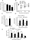
Intra-VTA microinjection of ibogaine decreases ethanol self-administration, and systemic ibogaine increases GDNF expression in a midbrain region that contains the VTA. A, B, Intra-VTA ibogaine decreased ethanol self-administration by rats. A, Ibogaine (0, 0.1, 1, 10 μm; equivalent to 0, 0.05, 0.5, 5.0 pmol) microinjected into the VTA 3 hr before an ethanol self-administration session dose-dependently decreased lever press responding (F(3,27) = 5.90; p < 0.002). Data are shown as mean ± SEM. **p < 0.001 compared with active lever responding after vehicle injection (n = 10). B, Time course of effect of intra-VTA ibogaine. The same subjects in A were tested 24 and 48 hr after ibogaine (IBO) treatment. There was no difference in responding on the day before treatment (Baseline) (F(3,27) = 0.36; p = 0.78). Ibogaine reduced responding for ethanol, and this reduction was long lasting (main effect of concentration: F(3,54) = 8.62, p < 0.001; main effect of time: F(2,54) = 6.76, p < 0.006; concentration × time interaction: F(6,54) = 0.26, p = 0.95). The 10 μm dose significantly reduced responding at all time points. The 0.1 μm dose was omitted for clarity; responding after this dose did not differ from vehicle at any tested time point. Data are shown as mean ± SEM. *p < 0.05, **p < 0.005 compared with active lever responding after vehicle injection (n = 10). C, Ibogaine microinjected into the substantia nigra 3 hr before an ethanol self-administration session did not affect ethanol lever press responding relative to vehicle microinjection (main effect of treatment: F(1,7) = 0.59, p = 0.47; main effect of lever: F(1,7) = 13.37, p < 0.007; treatment × lever interaction: F(1,7) = 0.38, P = 0.56). Data are shown as mean ± SEM (n = 8). D, E, The midbrain region was excised 1, 6, or 12 hr (mouse) and 1, 3, 24, and 48 hr (rat) after intraperitoneal injection of 40 mg/kg ibogaine. The expression of GDNF and control GPDH in mouse (D) and rat (E) was analyzed by RT-PCR. Histogram depicts the mean ratio (GDNF/GPDH) ± SD of n = 3 (D), n = 6 (1, 3, and 12 hr), and n = 5 (24 and 48 hr) (E). *p < 0.05, **p < 0.01 compared with saline injection.
Figure 3.
Figure 3.
Ibogaine is not neurotoxic to cells in culture. A-D, SHSY5Y cells were treated with vehicle (A), 10 μm ibogaine for 24 hr (B), 2 μm Latrunculin B (C), or the PI3 kinase inhibitor Wortmannin (4 μm) for 90 min (D). Cell death was measured with Fluoro-Jade as described in Materials and Methods.
Figure 4.
Figure 4.
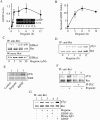
Ibogaine activates the GDNF pathway in SHSY5Y cells. A, Cells were treated with 10 μm ibogaine for the indicated times and lysed for total RNA isolation. Expression of GDNF and control actin was analyzed by RT-PCR (n = 4). B, Cells were treated with 10 μm ibogaine for the indicated times. GDNF in the media was detected by an ELISA assay. Histogram depicts the mean ± SD of GDNF secretion in three experiments. C, Cells were treated with the indicated concentrations of ibogaine for 3 hr. Ret was immunoprecipitated with anti-Ret antibodies, followed by Western blot analysis with anti-GFRα1 antibodies. The levels of GFRα1 in the homogenates were determined by Western blot analysis (n = 3). D, Cells were treated with 10 μm ibogaine for the indicated times. Ret was immunoprecipitated followed by Western blot analysis with anti-phosphotyrosine or anti-Ret antibodies (n = 3). E, Cells were treated with vehicle (lane1) or 10 μm ibogaine (lane 2) for 3 hr or with 50 ng/ml BDNF for 10 min (lane 3). Trk phosphorylation was analyzed by Western blot analysis with anti-phospho-Trk antibodies. The levels of TrkB were also determined by Western blot analysis (n = 3).F, Cells were preincubated with 0.3 U/ml PI-PLC for 1 hr (lanes 3 and 4). Cells were then washed and treated without (lanes 1 and 3) or with (lanes 2 and 4) 10 μm ibogaine for 3 hr. Ret phosphorylation was determined as described above (n = 4). G, Cells were treated for 3 hr with vehicle (lane 1), 10 μm ibogaine (lane 2), 10 μm ibogaine plus 10 μg/ml of mouse IgG (lane 3), or 10 μm ibogaine plus anti-GDNF neutralizing antibodies (lane 4). Treatment with 50 ng/ml GDNF was used as a positive control (lane 5). The cells were lysed and Ret phosphorylation was analyzed as described above (n = 3).
Figure 5.
Figure 5.
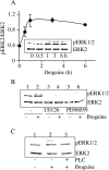
Ibogaine activates the MAPK signaling pathway. A, SHSY5Y cells were treated with 10 μm ibogaine for the indicated times. ERK2 and phosphoERK 1/2 (pERK1/2) were detected by Western blot analysis with anti-ERK2 and anti-pERK1/2 antibodies, respectively. Line graph depicts the mean ratio (pERK2/ERK2) ± SD of three experiments. B, Cells were preincubated with the inhibitors U0126 (20 μm) (lanes 3 and 4) and PD58089 (40 μm) (lanes 5 and 6) for 30 min and then treated without (lanes 1, 3, and 5) or with (lanes 2, 4, and 6) 10 μm ibogaine for 3 hr (n = 3). C, Cells were preincubated with 0.3 U/ml PI-PLC for 1 hr (lane 3). Cells were then washed and treated without (lanes 1) or with (lanes 2 and 3) 10 μm ibogaine for 3 hr (n = 3).
Figure 6.
Figure 6.
Intra-VTA infusion of GNDF mimics the effects of ibogaine, and anti-GDNF neutralizing antibodies attenuate the effects of ibogaine on ethanol self-administration. A, GDNF (5 μg/μl) microinjected into the VTA 10 min before an ethanol self-administration session decreased mean lever press responding relative to vehicle (main effect of treatment: F(1,7) = 8.38, p < 0.03; main effect of lever: F(1,7) = 17.10, p < 0.005; treatment × lever interaction: F(1,7) = 5.88, p < 0.05). **p < 0.003. The data are shown as mean lever presses ± SEM (n = 8). B, Rats received continuous infusion for 14 d of anti-GDNF neutralizing antibodies or mouse IgG (600 ng/12 μl per side per day) into the VTA via osmotic minipumps. Responding for ethanol reinforcement was measured before and after antibody infusion in daily 1 hr ethanol self-administration sessions. The effect of ibogaine (40 mg/kg, i.p.) was tested on the 10th day of antibody infusion, and self-administration behavior was measured for three additional daily sessions. “Pre-pump” represents the mean of the last three training sessions before antibody infusion; “Pre-Ibo” represents the mean of the last three training sessions after antibody infusion and before ibogaine injection. There was no effect of intra-VTA anti-GDNF antibody infusion on baseline levels of ethanol self-administration (Pre-pump vs Pre-Ibo) (F(1,15) = 0.244; p = 0.63). When the Pre-Ibo baseline was compared with data obtained 3 and 24 hr after ibogaine injection (Post-Ibo), there was a significant effect of antibody treatment (F(1,30) = 5.15; p < 0.04) and a significant effect of time (F(2,30) = 23.1; p < 0.001). Although ibogaine treatment decreased responding in both groups, the decrease in responding is significantly greater in the control mouse IgG group (n = 9) than in subjects treated with anti-GDNF neutralizing antibodies (n = 8). **p < 0.02; *p < 0.05. The data are shown as mean lever presses ± SEM.

Similar articles

Cited by

References

    1. Airaksinen MS, Saarma M (2002) The GDNF family: signaling, biological functions and therapeutic value. Nat Rev Neurosci 3: 383-394. - PubMed
    1. Alper KR, Lotsof HS, Frenken GM, Luciano DJ, Bastiaans J (1999) Treatment of acute opioid withdrawal with ibogaine. Am J Addict 8: 234-242. - PubMed
    1. Bailey CP, Manley SJ, Watson WP, Wonnacott S, Molleman A, Little HJ (1998) Chronic ethanol administration alters activity in ventral tegmental area neurons after cessation of withdrawal hyperexcitability. Brain Res 803: 144-152. - PubMed
    1. Bailey CP, O'Callaghan MJ, Croft AP, Manley SJ, Little HJ (2001) Alterations in mesolimbic dopamine function during the abstinence period following chronic ethanol consumption. Neuropharmacology 41: 989-999. - PubMed
    1. Balkowiec A, Katz DM (2000) Activity-dependent release of endogenous brain-derived neurotrophic factor from primary sensory neurons detected by ELISA in situ J Neurosci 20: 7417-7423. - PMC - PubMed

Publication types

MeSH terms