. 2005 Jan 19;25(3):637-47.
doi: 10.1523/JNEUROSCI.3960-04.2005.

Anti-CD11d integrin antibody treatment restores normal serotonergic projections to the dorsal, intermediate, and ventral horns of the injured spinal cord

Affiliations

Anti-CD11d integrin antibody treatment restores normal serotonergic projections to the dorsal, intermediate, and ventral horns of the injured spinal cord

Mark A Oatway et al. J Neurosci. .

Abstract

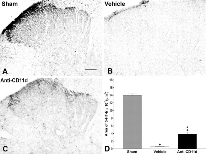
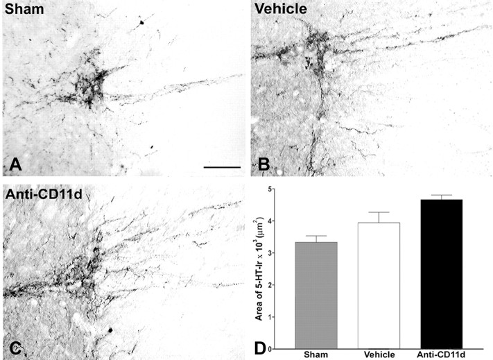
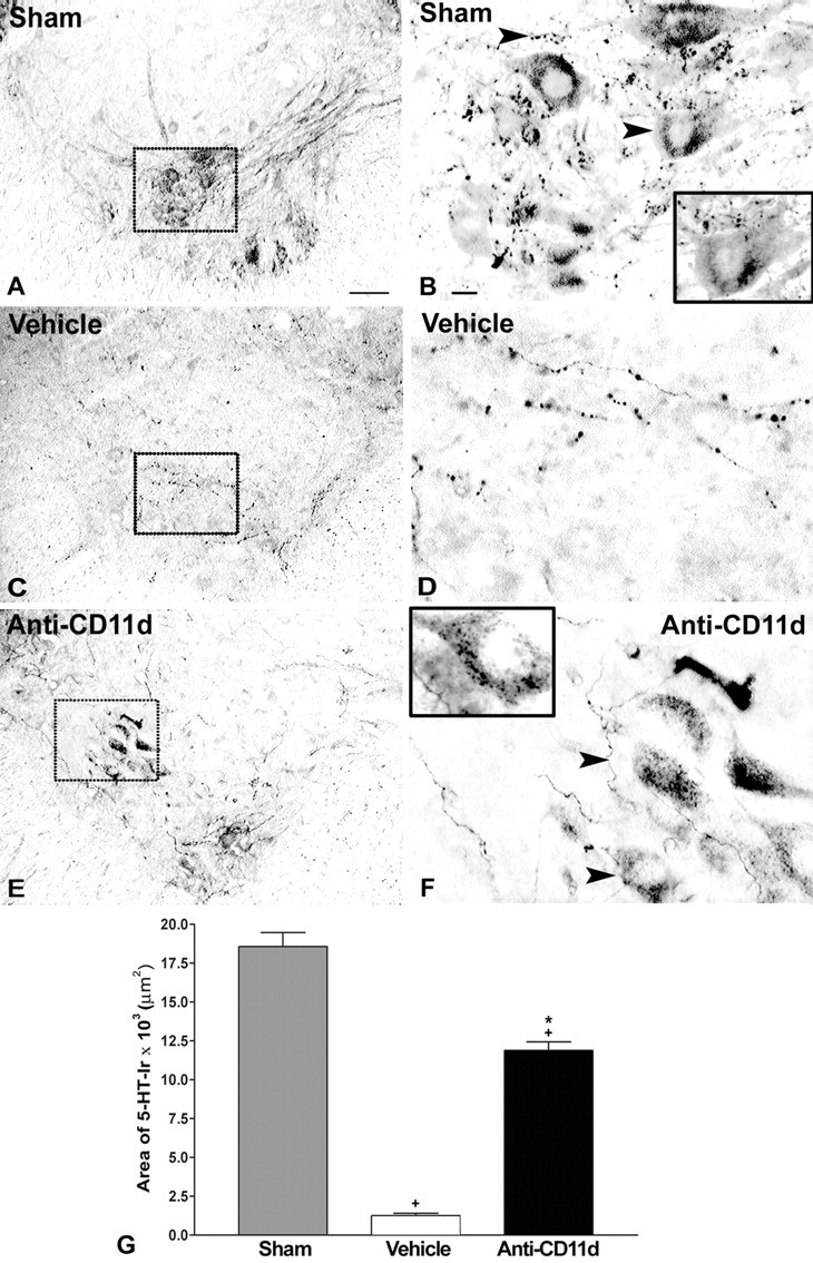
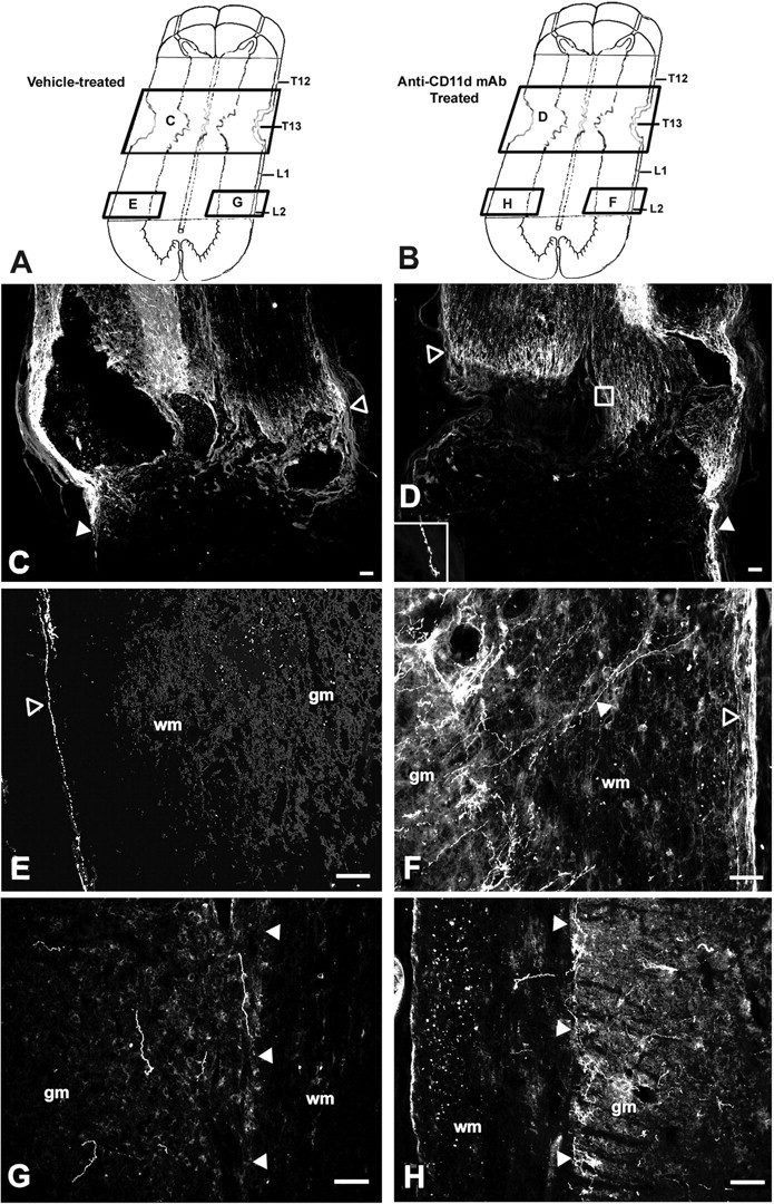
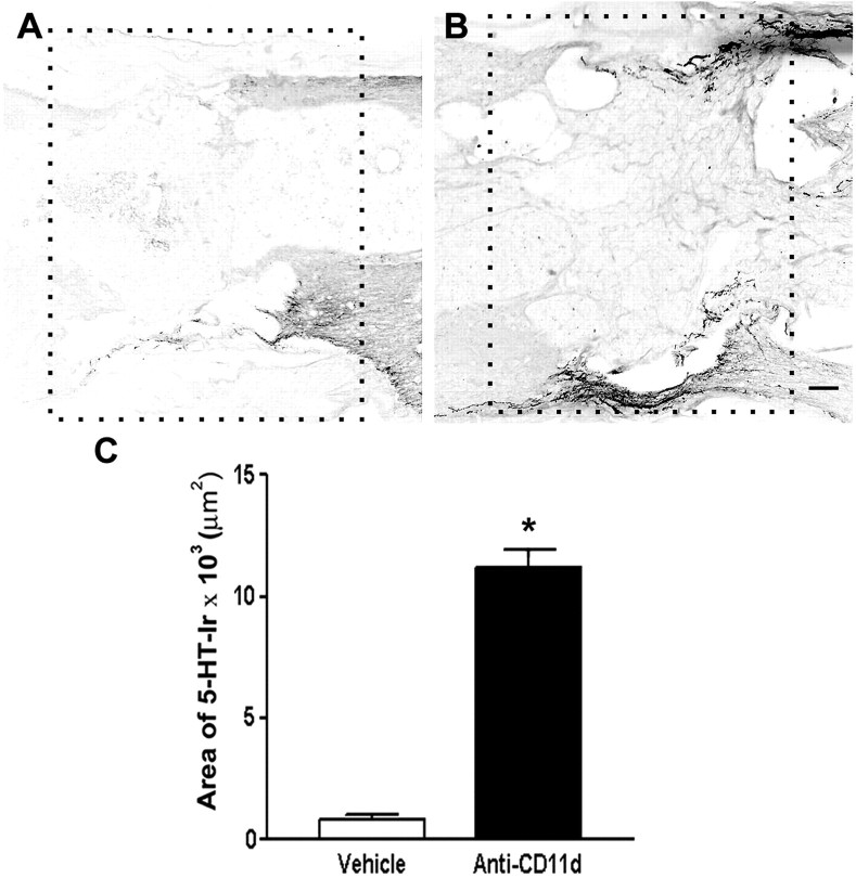
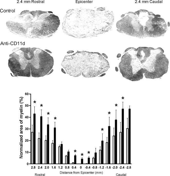
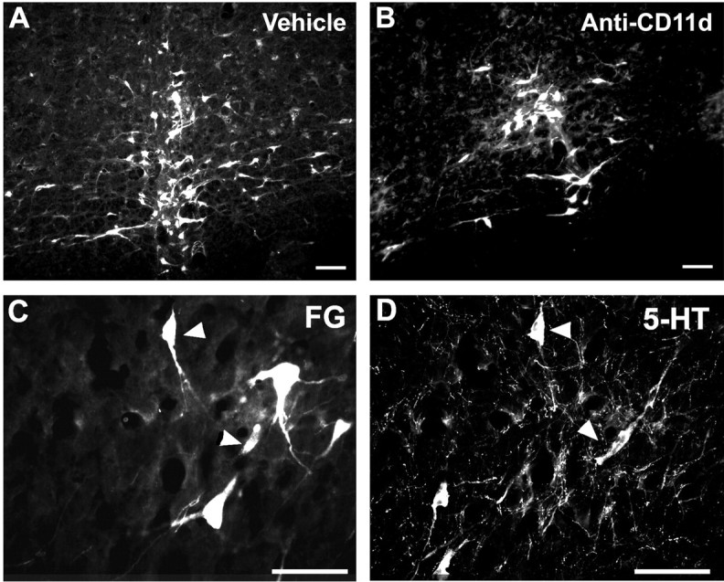
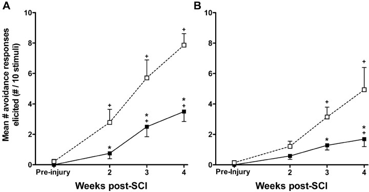
Spinal serotonergic pathways provide inhibitory and excitatory modulation of sensory, autonomic, and motor processing. After spinal cord injury (SCI), the acute inflammatory response is one process that damages descending pathways. Increases in serotonergic fiber density in spinal segments rostral and decreases caudal to the lesion have been observed previously and may contribute to neuropathic pain and motor dysfunction associated with SCI. We investigated the effect of an acute anti-inflammatory treatment on the density of serotonergic fibers rostral and caudal to a thoracic SCI lesion. This treatment, a monoclonal antibody to the CD11d subunit of the leukocyte CD11d/CD18 integrin, limits the trafficking of neutrophils and macrophages into the SCI site. In the dorsal horn, after treatment, the typically increased serotonin immunoreactivity rostral to injury was reduced, whereas that caudal to the lesion increased toward normal. Coincidently, mechanical allodynia in the dorsal trunk and hindpaws was significantly reduced. Increased serotonergic fiber density below the lesion also occurred in the intermediolateral cell column and ventral horn of treated rats, relative to controls. Improved locomotor recovery paralleled this increased serotonin. The treatment increased compact myelin in and near the lesion epicenter and increased serotonergic fiber bundles coursing around part of the lesion but had no consistent effect on the number of raphe-spinal neurons retrogradely labeled by tracer injection below the injury. In conclusion, this anti-CD11d integrin antibody treatment is neuroprotective after SCI, corresponding with improved patterns of intraspinal serotonergic innervation. The improvement in serotonergic fiber projections paralleled reduced mechanical allodynia and enhanced locomotor recovery.


Figures

Figure 1.
Figure 1.
5-HT-IR in laminas I-IV of the dorsal horn rostral to injury 4 weeks after SCI. Photomicrographs of transverse sections of the dorsal horn in sham-injured, vehicle-treated, and anti-CD11d mAb-treated animals. After SCI, the distribution and density of 5-HT-immunoreactive fibers were increased significantly, primarily in the superficial laminas, with punctate fibers in laminas III and IV. Anti-CD11d mAb treatment normalized the distribution of 5-HT-immunoreactive fibers toward patterns observed in sham-injured animals. Scale bar, 100 μm. The area of 5-HT-IR in the dorsal horn rostral to the injury at T12-13 represented the mean area ± SE of 5-HT-IR in the sham injured (n = 6), vehicle-treated (n = 5), and anti-CD11d mAb-treated (n = 6) groups. *p < 0.05 compared with vehicle-treated rats; +p < 0.05 compared with sham-injured rats.
Figure 2.
Figure 2.
5-HT-IR in laminas I-IV of the dorsal horn caudal to injury 4 weeks after SCI. Photomicrographs of transverse sections of the dorsal horn in sham-injured, vehicle-treated, and anti-CD11d mAb-treated animals. After SCI, 5-HT-immunoreactive fibers were completely absent in most dorsal horn sections. 5-HT-immunoreactive fibers were present as tortuous fiber within the superficial laminas after anti-CD11d mAb treatment. Scale bar, 100 μm. The area of 5-HT-IR (mean ± SE) in the dorsal horn caudal to the injury at T12-13 is plotted for the sham-injured (n = 5), vehicle-treated (n = 5), and anti-CD11d mAb-treated (n = 5) groups. *p < 0.05 compared with vehicle-treated rats; +p < 0.05 compared with sham-injured rats.
Figure 3.
Figure 3.
5-HT-IR in the intermediolateral cell column rostral to injury 4 weeks after SCI. Photomicrographs of transverse sections in the area of the intermediolateral cell column in sham-injured, vehicle-treated, and anti-CD11d mAb-treated animals. After SCI, the distribution and density of 5-HT-immunoreactive fibers were similar in all three groups. Scale bar, 50 μm. The area of 5-HT-IR (mean ± SE) in the intermediolateral cell column rostral to the injury at T12-13 is plotted for the sham-injured (n = 6), vehicle-treated (n = 5), and anti-CD11d mAb-treated (n = 6) groups.
Figure 4.
Figure 4.
5-HT-IR in the intermediolateral cell column caudal to injury 4 weeks after SCI. Photomicrographs of transverse sections of the intermediolateral cell column in sham-injured, vehicle-treated, and anti-CD11d mAb-treated animals. After SCI, the varicose, 5-HT-immunoreactive fibers were sparsely distributed within the IML. Anti-CD11d mAb treatment resulted in approximately a twofold increase in 5-HT-IR characterized by a robust fiber distribution and rounded, somata-like structures. Scale bar, 50 μm. The area of 5-HT-IR (mean ± SE) in the intermediolateral cell column caudal to the injury at T12-13 is plotted for the sham-injured (n = 6), vehicle-treated (n = 5), and anti-CD11d mAb-treated (n = 6) groups. *p < 0.05 compared with vehicle-treated rats; +p < 0.05 compared with sham-injured rats.
Figure 5.
Figure 5.
5-HT-IR in laminas VII-IX of the ventral horn caudal to injury 4 weeks after SCI. A-F, Photomicrographs of transverse sections of the ventral horn in sham-injured (A, B), vehicle-treated (C, D), and anti-CD11d mAb-treated (E, F) animals. After SCI, the distribution and density of 5-HT-immunoreactive fibers were significantly reduced to sparsely distributed varicose fibers observed in control animals (C, D). Anti-CD11d mAb treatment resulted in increased distribution of 5-HT-IR in fibers and terminal boutons (arrowheads) opposing the somata of α-motoneurons (E,F), similar to patterns observed in sham-injured animals (A,B). B,G, Insets, Higher-power photomicrographs of the neuron indicated by an arrow in this field of view. Scalebars: A,C,E, 100 μm; B,D,F, 20 μm. G, The area of 5-HT-IR in the ventral horn caudal to the lesion site represented as the mean area ± SE of 5-HT-IR in the sham-injured (n = 5), vehicle-treated (n = 6), and anti-CD11d mAb-treated (n = 5) groups. *p < 0.05 compared with vehicle-treated rats; +p < 0.05 compared with sham-injured rats.
Figure 6.
Figure 6.
Digital photomicrographs demonstrating the presence of 5-HT-immunoreactive fibers around and caudal to the T12-L1 lesion in vehicle-treated (C, E, G) and anti-CD11d mAb-treated (D, F, H) animals. Locations of each photomicrograph, with respect to the lesion site, are depicted in A for vehicle-treated and in B for anti-CD11d mAb-treated rats. Longitudinal sections in vehicle-treated animals (n = 5) revealed 5-HT-immunoreactive fibers at the rostral border of the lesion (C, open arrowhead) unable to penetrate in to the scar. 5-HT-immunoreactive fibers were observed unilaterally, descending along the subpial rim of the cord ∼3.5 mm caudal to the lesion border (E, open arrowhead). Individual 5-HT-immunoreactive fibers were distributed sparsely within the gray matter caudal to the lesion in randomized patterns, with no apparent connection to the descending fibers (G). Longitudinal sections in anti-CD11 mAb-treated animals (n = 4) revealed 5-HT-IR at the rostral border of the lesion (D, open arrowhead), with characteristic axonal retraction (D, inset) unable to permeate the scar. Fibersal so extended caudally through the lesion along the subpial rim. Approximately 3.5 mm caudal to the lesion site, collateral axons extended medially from the subpial rim toward the gray matter border (designated by filled arrowhead) where 5-HT-immunoreactive fibers accumulated densely within the IML (F). On the contralateral side of the cord, 5-HT-immunoreactive fibers were present within the IML, in the absence of fiber connections from the contralateral or ipsilateral pial rim (H). gm, Gray matter; wm, white matter. Scale bars: C, D, 100 μm; E-H, 50 μm.
Figure 7.
Figure 7.
Serotonergic fiber density in and adjacent to the lesion site in vehicle-treated (n= 4) and anti-CD11d mAb-treated (n = 3) rats. Low-power photomicrographs of the lesion areas analyzed in the vehicle- and mAb-treated rats are outlined with boxes in A and B, respectively. The rostral side of lesion is on the right of photomicrographs. Images have been reversed to black on white to reveal details of lesion structure. Scale bar, 200 μm. C, Graph illustrates mean area of immunoreactivity for 5-HT within the lesion in vehicle- and mAb-treated rats. Data are expressed as mean values ± SEM. *p < 0.05 compared with vehicle-treated rats.
Figure 8.
Figure 8.
Normalized area of spared myelin stained with Luxol fast blue within the lesion site after SCI. Photomicrographs illustrate compact myelin integrity 2.4 mm rostral to epicenter, within the epicenter, and 2.4 mm caudal to epicenter in a vehicle-treated and an anti-CD11d-treated rat. Anti-CD11d-treated animals (filled bars) (n = 4) had significantly increased amounts of compact myelin at the epicenter and in most rostral areas compared with vehicle-treated animals (open bars) (n = 4). *p < 0.05 compared with vehicle-treated rats.
Figure 9.
Figure 9.
Fluorogold-labeled neurons in the nucleus raphe magnus and nucleus raphe pallidus of vehicle-treated (n = 6) (A) and anti-CD11d mAb-treated (n = 5) (B) animals. Many of these neurons were immunoreactive for 5-HT (C, D, arrowheads indicate double-labeled cells). Scale bars: A, B, 100 μm; C, D, 50 μm.
Figure 10.
Figure 10.
The development of mechanical allodynia at the level (A) and below the level (B) of SCI lesion site. Testing sessions consisted of 10 stimulations with an innocuous, monofilament stimulus 1 week before injury and at 2, 3, and 4 weeks after injury. Each data point represents the mean ± SEM number of avoidance responses made to 10 stimulations. A, The number of avoidances made in response to dorsal trunk stimulation was significantly reduced after anti-CD11d mAb treatment (filled squares) (n = 7) when compared with vehicle-treated animals (open squares) (n = 8), suggesting the reduction of at-level mechanical allodynia. B, The number of paw withdrawals was also reduced in the anti-CD11d mAb-treated group (filled squares) (n = 7) when compared with the vehicle-treated group (open squares) (n = 8), suggesting attenuation of below-level mechanical allodynia. *p < 0.05 compared with vehicle-treated rats; +p < 0.05 compared with mean responses before injury.
Figure 11.
Figure 11.
BBB open-field locomotor scores assessed from 5 d to 4 weeks after SCI. Mean ± SEM scores were generated as an average of the individual scores from the two hindlimbs. CD11d mAb-treated rats (filled squares) (n = 9) had significantly higher locomotor scores at days 8, 15, 19, 22, and 26 compared with vehicle-treated rats (open squares) (n=7). *p < 0.05 compared with vehicle-treated rats.

Similar articles

Cited by

References

    1. Ainge G, Cook JL, Gisby PT (1969) Rapid staining of myelin in paraffin sections with Luxol fast blue MBS. J Med Lab Technol 26: 231-232. - PubMed
    1. Ali Z, Wu G, Kozlov A, Barasi S (1996) The role of 5HT3 in nociceptive processing in the rat spinal cord: results from behavioural and electrophysiological studies. Neurosci Lett 208: 203-207. - PubMed
    1. Allen GV, Cechetto DF (1994) Serotoninergic and nonserotoninergic neurons in the medullary raphe system have axon collateral projections to autonomic and somatic cell groups in the medulla and spinal cord. J Comp Neurol 350: 357-366. - PubMed
    1. Bao F, Chen Y, Dekaban GA, Weaver LC (2004a) An anti-CD11d integrin antibody reduces cyclooxygenase-2 expression and protein and DNA oxidation after spinal cord injury in rats. J Neurochem 90: 1194-1204. - PubMed
    1. Bao F, Chen Y, Dekaban GA, Weaver LC (2004b) Early anti-inflammatory treatment reduces lipid peroxidation and protein nitration after spinal cord injury in rats. J Neurochem 88: 1335-1344. - PubMed

Publication types

MeSH terms