. 2005 Jan 19;25(3):737-47.
doi: 10.1523/JNEUROSCI.4174-04.2005.

Expression of stathmin, a developmentally controlled cytoskeleton-regulating molecule, in demyelinating disorders

Affiliations

Expression of stathmin, a developmentally controlled cytoskeleton-regulating molecule, in demyelinating disorders

Aixiao Liu et al. J Neurosci. .

Abstract

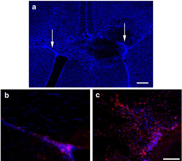
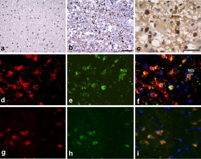
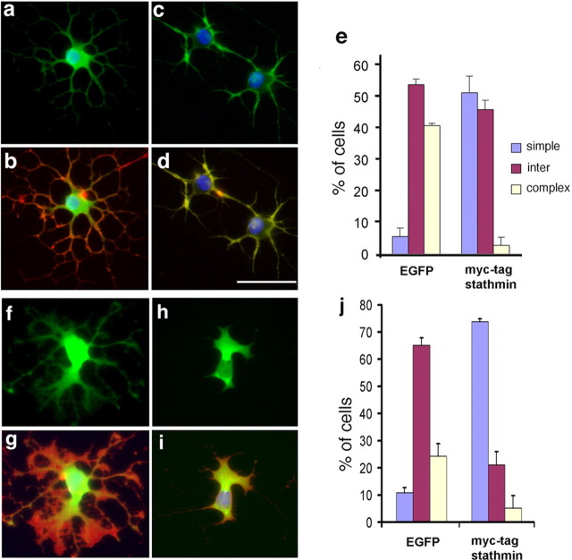
Understanding the biological relevance of reexpression of developmental molecules in pathological conditions is crucial for the development of new therapies. In this study, we report the increased expression of stathmin, a developmentally regulated tubulin-binding protein, in the brains of patients with multiple sclerosis (MS). In physiological conditions, stathmin immunoreactivity was observed in polysialic acid-neural cell adhesion molecule-positive migratory progenitors in the subventricular zone, and its expression progressively decreased as the cells matured into oligodendrocytes (OLs). In MS patients, however, stathmin levels were elevated in 2',3'-cyclic nucleotide 3'-phosphodiesterase-positive OLs, in 10 of 10 bioptic samples analyzed. Increased levels of stathmin were confirmed by Western blot analysis of normal-appearing white matter samples from MS brains. In addition, using mass spectrometry, stathmin was identified as the main component of a specific myelin protein fraction consistently increased in MS preparations compared with controls. To test the biological relevance of increased stathmin levels, primary OL progenitors were transfected using a myc-tagged stathmin cDNA and were allowed to differentiate. Consistent with a distinct role played by this molecule in cells of the OL lineage at different developmental stages, transient transfection in progenitors favored the bipolar migratory phenotype but did not affect survival. However, sustained stathmin levels in differentiating OLs, because of overexpression, resulted in enhanced apoptotic susceptibility. We conclude that stathmin expression in demyelinating disorders could have a dual role. On one hand, by favoring the migratory phenotype of progenitors, it may promote myelin repair. On the other hand, stathmin in mature OLs may indicate cell stress and possibly affect survival.


Figures

Figure 1.
Figure 1.
Progressive decrease of stathmin levels during oligodendrocyte morphological differentiation in vitro. Immunofluorescent staining of stathmin (red) in neonatal primary rat cortical progenitors cultured on poly-lysine-coated slides (a-f) and maintained undifferentiated in medium containing PDGF plus bFGF (a, b) or differentiated by mitogen withdrawal (c, d) or by PDGF plus T3 treatment (e, f) for 3 d are shown. Morphological differentiation and myelin membrane formation was also assessed by culturing cells on laminin-coated slides (g-j) in the presence of PDGF plus T3 (g, h) or in the absence of mitogens (i, j). Undifferentiated bipolar progenitors were identified by positive immunoreactivity for the early oligodendrocyte progenitor marker A2B5 (green; b), whereas branched developing oligodendrocytes were identified by O4 immunoreactivity (green; d, f, h, j). Note that stathmin immunoreactivity (red; a-j) was reduced during differentiation in vitro and that growth on laminin further decreased its levels and confined its localization to the soma of the differentiated cells (g, i) with complete exclusion from the cytoplasmic processes and the myelin membrane (j). Scale bar, 40 μm. Phosphorylated stathmin was detected by Western blot analysis (k) using antibodies generated against specific phosphorylated serine residues, such as serine 16 (S16), serine 25 (S25), and serine 38 (S38). Note that stathmin was weakly phosphorylated on residues 25 and 38. Actin was used as a loading control.
Figure 2.
Figure 2.
Stathmin is expressed in cells of the SVZ and in the corpus callosum. Confocal images (a-l) of the mouse corpus callosum (CC) and SVZ stained with stathmin (red) and with antibodies specific for the migratory SVZ progenitors PSA-NCAM (green; b, c), the oligodendrocytic marker APC/CC1 (green; e, f), the astrocytic marker GFAP (green; h, i), and the axonal marker neurofilament NF-M (green; k, l) are shown. DAPI (blue) was used as a nuclear counterstain. Note that intense stathmin immunoreactivity was observed only in the SVZ (a, d, g, j) and in sporadic cells in the adult corpus callosum. Stathmin-positive cells were often CC1 negative (e), with the exception of few weakly positive cells (e, white arrows) that could be better appreciated at a high-power view of the same field (f). Note that stathmin immunoreactivity did not colocalize with the astrocytic marker GFAP (g-i) or with the axonal marker neurofilament NF-M (j-l). In contrast, the vast majority of the stathmin-positive cells in the SVZ were PSA-NCAM positive (a-c). Scale bar, 50 μm.
Figure 3.
Figure 3.
Stathmin expression in SVZ migratory progenitors is increased in response to chemically induced demyelination. Demyelination was induced by ethidium bromide injection in the corpus callosum on the right side. Four days after injection, mice were perfused, and the brains were removed and cryosectioned. DAPI staining (blue) was performed to detect the nuclei of the cells and to reveal cell loss on the injected side (a, right arrow) compared with the noninjected side (a, left arrow). Immunofluorescence staining using antibodies against stathmin (red; b, c) revealed a larger number of stathmin-positive cells in the SVZ and in the region surrounding the lesion site (c) compared with the noninjected side (b). Scale bars: a, 100 μm; b, c,50 μm.
Figure 4.
Figure 4.
Stathmin expression decreases as progenitor cells mature into oligodendrocytes to repair the lesion caused by ethidium bromide injection. Four days after ethidium bromide injection, stathmin-positive cells (red; a, b, d, e) were colabeled by antibodies recognizing the oligodendrocyte progenitor marker Sox10 (green; a, c) and PSA-NCAM (green; d, f). Although several of the stathmin-positive cells were Sox10 positive (a-c, arrow), most of the stathmin-positive cells were identified by the marker for migratory progenitors PSA-NCAM (d-f, arrows). Two weeks after injection, when the process of remyelination begins, the newly generated mature oligodendrocytes identified by APC/CC1 staining (green; g, i) did not show stathmin immunoreactivity (g-i). Scale bar, 50 μm.
Figure 5.
Figure 5.
Increased stathmin levels in the brain of MS patients. Very few immunoreactive cells (dark brown staining) were found in the white matter of control brain sections counterstained with hematoxylin (purple staining in a). In contrast, numerous stathmin-immunoreactive oligodendrocyte-like cells (dark brown staining) were found in the highly inflamed lesional area in MS patients (b; original magnification, 400×). Stathmin (brown staining) was found predominantly in the soma and in the processes of oligodendrocytes in MS lesions counterstained with hematoxylin (c; original magnification, 1000×). Note that no stathmin immunoreactivity was observed in axons (c). Immunofluorescence of MS brain lesions using anti-CNPase (red; d) and stathmin (green; e) antibodies further confirmed that the immunoreactive cells belong to the oligodendrocyte lineage, as seen in the overlay of the two panels (f). The stathmin-positive cells (green; h) in MS brain lesions are also APC/CC1 immunoreactive (red; g). Double-stained cells appear yellow (i).
Figure 6.
Figure 6.
Increased stathmin protein levels in the brain of MS patients. To characterize the expression of stathmin in the human normal-appearing white matter from patients (MS) and normal controls (control), we adopted a fractionation method described previously for the isolation of basic proteins (Wood and Moscarello, 1989). Several fractions named C1 to C8a and C8b were isolated from the white matter samples of control and MS human brains (a). The yield of the C1 to C3 fractions was dramatically decreased in MS patients, whereas fractions C8a and C8b were increased compared with controls (b). A Coomassie blue staining of equal amount of proteins isolated from these different fractions and separated by gel electrophoresis (b, coomassie) revealed the presence of prominent bands in the 17-18 kDa molecular weight range in each fraction (double arrows). Mass spectrometry confirmed that MBP and its variously charged isomers were the main component of the 17-18 kDa band in fractions C1 to C4 and C8b, whereas stathmin was identified with a high level of confidence (individual ion score of >33) as the main band in fraction C8a. Each experiment was performed on samples from three normal and four patients and performed in triplicate. Western blot analysis with antibodies against MBP or stathmin confirmed the mass spectrometry results (b). Note that stathmin was identified as a doublet with the lower band significantly increased in MS patients. Western blot analysis revealed a similar increase in the stathmin doublet also in whole-cell lysate preparations obtained from the normal-appearing white matter of MS patients compared with age- and sex-matched samples. Actin was used as loading control (c). Quantitative slot-immunoblot analysis of the acid-extracted white matter C8 fractions isolated from control and MS brain samples further confirmed the increased stathmin levels in MS brain compared with control (d). Quantification of these results is shown as a bar graph (e). Although some variability was observed for the various patients, stathmin levels in the membrane fractions isolated from MS brain tissue were almost twice as high as the levels found in the control brains. The effect of inflammatory cytokines on stathmin expression in the oligodendrocyte lineage was therefore tested in vitro, by treating differentiating oligodendrocyte progenitors (OL) with 0.2 μg/ml TGF-β (OL plus TGF-β) or TNF-α (OL plus TNF-α) for 24 h after mitogen withdrawal. f, Cytokine treatment of primary cultured cells resulted in increased levels of stathmin mRNA, as detected by RT-PCR, and protein, as detected by Western blot analysis. Actin was used as a control for equal loading.
Figure 7.
Figure 7.
Stathmin gain-of-function in progenitors prevents the attainment of a highly branched phenotype. Immunoselected A2B5-positive rat cortical oligodendrocyte progenitors were electroporated with a mammalian expression vector containing either EGFP (a, b, f, g) or myc-tagged stathmin (c, d, h, i) and then plated on poly-lysine-coated (a-d) or laminin-coated (f-i) chamber slides. The day after transfection, cells were induced to differentiate in mitogen-free chemically defined medium for 24 h and then fixed. Double immunofluorescence for O4 (red; b, d, g, i) and either GFP (green; a, b, f, g) or myc-tag (green; c, d, h, i) was used to identify the morphology of the transfected cells. DAPI (blue; a-d, f-i) was used as a nuclear counterstain. A total of >500 transfected cells from triplicate experiments were classified according to their morphology as simple, intermediate, or complex and then quantified (e, j). Stathmin overexpression increased cells with simple and intermediate morphology and prevented the attainment of a complex phenotype. Note that this inhibitory effect on branching occurred independently of the substrate. The difference between the number of cells with simple morphology in the GFP-transfected versus the myc-stathmin-transfected groups was statistically significant (p < 0.005). Scale bar, 100 μm.
Figure 8.
Figure 8.
Elevated levels of stathmin in differentiating, but not in proliferating, oligodendrocyte progenitors enhance the susceptibility of these cells to apoptotic stimuli. Myc-tagged stathmin- and EGFP-transfected progenitors kept in PDGF plus bFGF (a) or allowed to differentiate for 3 d by mitogen removal (b) were subject to glucose deprivation during the last 24 h, and the number of surviving transfectants was calculated. Note that high levels of stathmin did not affect survival of proliferating progenitors (a), whereas it enhanced the susceptibility of differentiating cells to death (b). To confirm that in stathmin-overexpressing cells were apoptotic, TUNEL+ cells (green; c, d) were counted in pcDNA-transfected (c) and myc-stathmin-transfected (d) cells. The percentage of TUNEL + cells was calculated by referring the number to the total number of DAPI + cells (blue; c, d), and the data are presented as a bar graph (e). The difference in apoptotic cells between the two groups of differentiating oligodendrocytes was statistically significant (p < 0.005). Scale bar, 100 μm.

Similar articles

Cited by

References

    1. Ainger K, Avossa D, Morgan F, Hill SJ, Barry C, Barbarese E, Carson JH (1993) Transport and localization of exogenous myelin basic protein mRNA microinjected into oligodendrocytes. J Cell Biol 123: 431-441. - PMC - PubMed
    1. Almeida A, Delgado-Esteban M, Bolanos JP, Medina JM (2002) Oxygen and glucose deprivation induces mitochondrial dysfunction and oxidative stress in neurones but not in astrocytes in primaryculture. J Neurochem 81: 207-217. - PubMed
    1. Amat JA, Fields KL, Schubart UK (1991) Distribution of phosphoprotein p19 in rat brain during ontogeny: stage-specific expression in neurons and glia. Dev Brain Res 60: 205-218. - PubMed
    1. Anderson DJ, Axel R (1985) Molecular probes for the development and plasticity of neural crest derivatives. Cell 42: 649-662. - PubMed
    1. Bakshi R, Miletich RS, Kinkel PR, Emmet ML, Kinkel WR (1998) Highresolution fluorodeoxyglucose positron emission tomography shows both global and regional cerebral hypometabolism in multiple sclerosis. J Neuroimaging 8: 228-234. - PubMed

Publication types

MeSH terms

LinkOut - more resources