. 2005 Jan 19;25(3):758-67.
doi: 10.1523/JNEUROSCI.3909-04.2005.

Neurotrophin-3 suppresses thermal hyperalgesia associated with neuropathic pain and attenuates transient receptor potential vanilloid receptor-1 expression in adult sensory neurons

Affiliations

Neurotrophin-3 suppresses thermal hyperalgesia associated with neuropathic pain and attenuates transient receptor potential vanilloid receptor-1 expression in adult sensory neurons

Tracy D Wilson-Gerwing et al. J Neurosci. .

Abstract

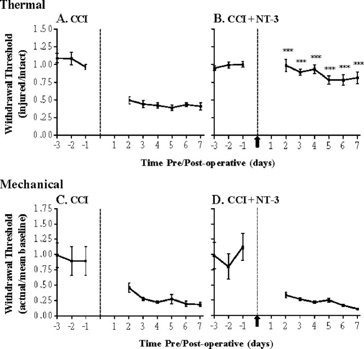
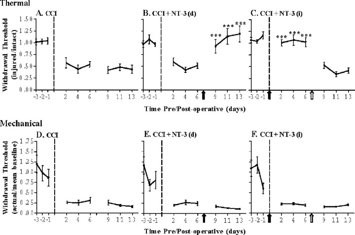
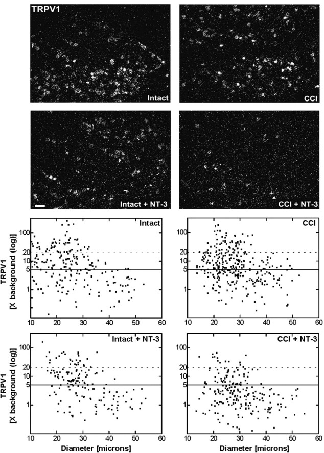
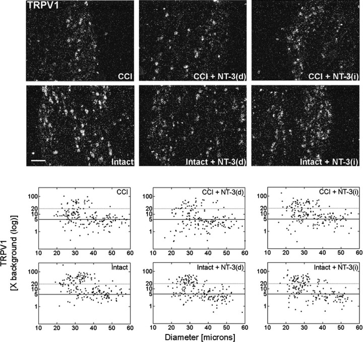
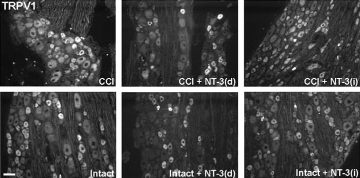
Neurotrophin-3 (NT-3) negatively modulates nerve growth factor (NGF) receptor expression and associated nociceptive phenotype in intact neurons, suggesting a beneficial role in treating aspects of neuropathic pain mediated by NGF. We report that NT-3 is effective at suppressing thermal hyperalgesia associated with chronic constriction injury (CCI); however, NT-3 does not alter the mechanical hypersensitivity that also develops with CCI. Thermal hyperalgesia is critically linked to expression and activation of the capsaicin receptor, transient receptor potential vanilloid receptor-1 (TRPV1). Thus, its modulation by NT-3 after CCI was examined. CCI results in elevated TRPV1 expression at both the mRNA and protein levels in predominantly small-to-medium neurons, with the percentage of neurons expressing TRPV1 remaining unchanged at approximately 56%. Attenuation of thermal hyperalgesia mediated by NT-3 correlates with decreased TRPV1 expression such that only approximately 26% of neurons ipsilateral to CCI expressed detectable TRPV1 mRNA. NT-3 effected a decrease in expression of the activated component of the signaling pathway linked to regulation of TRPV1 expression, phospho-p38 MAPK (Ji et al., 2002), in neurons ipsilateral to CCI. Exogenous NT-3 could both prevent the onset of thermal hyperalgesia and reverse established thermal hyperalgesia and elevated TRPV1 expression 1 week after CCI. Continuous infusion is required for suppression of both thermal hyperalgesia and TRPV1 expression, because removal of NT-3 resulted in a prompt reestablishment of the hyperalgesic state and corresponding CCI-associated TRPV1 phenotype. In conclusion, although NGF drives inflammation-associated thermal hyperalgesia via its regulation of TRPV1 expression, NT-3 is now identified as a potent negative modulator of this state.


Figures

Figure 1.
Figure 1.
NT-3 prevents the development of thermal hyperalgesia but not mechanical sensitivity associated with CCI. Hyperalgesic index plots represent alterations in thermal (A, B) and mechanical (C, D) sensitivities in response to 7 d CCI without (A, C) or with (B, D) 7 d NT-3 intrathecal infusion. Dashed line indicates the time of unilateral CCI. A, Thermal hyperalgesia is significantly increased compared with baseline levels after CCI and is maintained for the duration of the experiment (n = 7). B, Infusion of NT-3 significantly attenuates the development of thermal hyperalgesia after CCI and is maintained for the duration of the infusion (***p < 0.0001, relative to CCI alone; n = 5). The solid arrow indicates the start of NT-3 infusion. C, Mechanical hypersensitivity is significantly increased compared with baseline levels after unilateral CCI and is maintained for the duration of the experiment (7 d; n = 7). D, Infusion of NT-3 does not alter the mechanical hypersensitivity that develops after unilateral CCI (7 d; n = 5). The solid arrow indicates the start of NT-3 infusion.
Figure 2.
Figure 2.
NT-3 does not significantly alter thermal or mechanical thresholds in naive animals. Hyperalgesic index plots represent alterations in thermal (A-C) and mechanical (D-F) sensitivities in naive animals in response to vehicle (B, E) or NT-3 (C, F) intrathecal infusion for 7 d. A, Naive, uninjured animals demonstrate little fluctuation in their responses to thermal stimuli compared with baseline (n = 5). B, Infusion of control (1× vehicle) did not alter responses to thermal stimuli in naive animals compared with baseline (n = 3). The arrow indicates the time of infusion of control. C, Infusion of NT-3 did not alter responses to thermal stimuli in naive animals compared with baseline levels (n = 6). The solid arrow indicates the time of infusion of NT-3. D, Naive, uninjured animals do not demonstrate significant fluctuation in their responses to mechanical stimuli compared with baseline (n = 5). E, Infusion of control (1× vehicle) did not significantly alter responses to mechanical stimuli in naive animals compared with baseline levels (n = 3). The arrow indicates the time of infusion of vehicle control. F, Infusion of NT-3 did not significantly alter responses to mechanical stimuli in naive animals compared with baseline levels; however, a nonsignificant increased mechanical sensitivity was observed (n = 6). The solid arrow indicates the start of NT-3 infusion.
Figure 3.
Figure 3.
Delayed infusion of NT-3 can reverse established thermal hyperalgesia but not mechanical sensitivity after CCI. Hyperalgesic index plots represent alterations in thermal (A-C) and mechanical(D-F) sensitivities in 14 dCCI animals in response to delayed NT-3 [7 d post-CCI, (B, E)] or immediate NT-3 [followed by removal for the last 7 d (C, F)] intrathecal infusion. Dashed line indicates the time of CCI; solid arrow indicates the start of NT-3 infusion; open arrow indicates the time of pump removal. A, Thermal hyperalgesia is significantly increased compared with baseline levels after unilateral CCI and is maintained for the duration of the experiment (n = 7). B, Delayed infusion of NT-3 significantly reverses thermal hyperalgesia compared with CCI alone (***p < 0.0001) and is maintained for the duration of the infusion (7 d) (n = 7). Asterisks indicate significant difference from CCI alone. C, Infusion of NT-3 is required for continuous prevention of thermal hyperalgesia after CCI. Infusion of NT-3 significantly attenuates the development of thermal hyperalgesia after unilateral CCI and is maintained for the duration of the 7 d infusion (***p < 0.0001). After removal of the NT-3 pump, thermal hyperalgesia was reestablished and did not significantly differ from CCI alone (n = 7). The solid arrow indicates the start of NT-3 infusion; open arrow indicates the time of pump removal. Asterisks indicate significant difference from CCI alone. D, Mechanical sensitivity is significantly increased compared with baseline levels after unilateral CCI and is maintained for the duration of the experiment (n = 7). E, Delayed infusion of NT-3 does not alter mechanical sensitivity compared with CCI alone (n = 7). F, Stopping NT-3 infusion does not alter mechanical sensitivity compared with CCI alone (n = 7).
Figure 4.
Figure 4.
Message levels for TRPV1 are reduced after NT-3 treatment. Top, Dark-field photomicrographs of 6-μm-thick adult rat L5 DRG sections processed for in situ hybridization to detect TRPV1 transcripts contralateral (intact) or ipsilateral to 7 d CCI (CCI) and after a 7 d unilateral CCI plus intrathecal infusion of 600 ng · μl-1 · h-1 NT-3 (Intact + NT-3; CCI + NT-3). Scale bar, 100 μm. Note: NT-3 infusion results in the reduction in relative levels of hybridization signal for TRPV1 over individual neurons, with the influence most apparent after CCI. Bottom, Representative scatterplots whereby each point represents the labeling index of an individual neuron identified in 6-μm-thick sections of L5 DRG processed to detect TRPV1 mRNA. The relationship between TRPV1 mRNA labeling intensity (y-axis, log scale) and perikaryal diameter (x-axis) is depicted. Experimental states are indicated at the top right of each graph and are as described above. Labeling refers to the ratio of silver grain density over the neuronal cytoplasm to grain density over areas of the neuropil devoid of positive hybridization signal. Solid lines divide the plots into labeled and unlabeled populations; dotted lines separate lightly labeled from moderate to heavily labeled populations of TRPV1-expressing neurons. Note: In DRG contralateral to CCI, TRPV1 is expressed predominantly in small-to-medium neurons. CCI results in elevated TRPV1 expression in small-to-medium neurons and a novel but low level of expression in large neurons. NT-3 infusion produced a reduction in the levels and percentage of neurons expressing detectable TRPV1 mRNA DRG both ipsilateral and contralateral to CCI, with a more pronounced effect ipsilateral to injury.
Figure 5.
Figure 5.
Delayed NT-3 infusion reverses the increase in TRPV1 mRNA detected in DRG neurons subjected to CCI. Top, Dark-field photomicrographs of 6 μm sections of L5 DRG ipsilateral or contralateral to 14 d unilateral CCI, in response to 7 d 600 ng · μl-1 · h-1 NT-3 infusion for the last 7 d of a 14 d unilateral CCI [CCI + NT-3(d); Intact + NT-3(d)], or in response to 7 d 600 ng · μl-1 · h-1 NT-3 infusion for the first 7 d of a 14 d unilateral CCI [CCI + NT-3(i); Intact + NT-3(i)], as indicated. Scale bar, 60 μm. Note: Delayed infusion of NT-3 [CCI + NT-3(d)] results in a decrease in the relative levels of TRPV1 mRNA detected. Bottom, Representative scatterplots whereby each point represents the labeling index of an individual neuron identified in 6-μm-thick sections of L5 DRG processed to detect TRPV1 mRNA. The relationship between TRPV1 mRNA labeling intensity (y-axis, log scale) and perikaryal diameter (x-axis) is depicted. Experimental states as indicated are as described above. Labeling refers to the ratio of silver grain density over the neuronal cytoplasm to grain density over areas of the neuropil devoid of positive hybridization signal. Solid lines divide the plots into labeled and unlabeled populations; dotted lines separate lightly labeled from moderate to heavily labeled populations of TRPV1-expressing neurons. Note: Delayed infusion of NT-3 [CCI + NT-3(d)] results in a decrease in the relative levels of TRPV1 most prominent in medium-to-large neurons.
Figure 6.
Figure 6.
NT-3 infusion results in decreased expression of TRPV1 protein. Top, Fluorescence photomicrographs demonstrate levels of TRPV1 and p-p38 MAPK-like immunoreactivity in 10 μm sections of DRG ipsilateral (CCI) and contralateral to CCI (Intact) L5 DRG with or without immediate intrathecal infusion of NT-3 (CCI + NT-3, Intact + NT-3), as indicated. Scale bar, 60 μm. Note: In the DRG contralateral to CCI (Intact), levels of TRPV1 protein are highest in small neurons, with lower levels of expression observed in several small-to-medium dorsal root ganglion neurons. Seven days after CCI, levels of expression have increased relative to the intact state, with protein now being detected in a few large neurons. Intrathecal infusion of NT-3 at the time of injury results in reduced levels of TRPV1 protein, most notable in medium-to-large CCI neurons. Levels of p-p38 MAPK nuclear expression do not appear to be altered by CCI. Cytoplasmic expression of p-p38 MAPK does decrease slightly with CCI. Infusion of NT-3 results in a dramatic downregulation of p-p38 MAPK after CCI. The DRG contralateral to CCI (Intact) appear to have an increase in perineuronal p-p38 MAPK staining. Bottom, Representative Western blots of TRPV1 (n = 4) and p-p38 MAPK (n = 5) reflect the staining patterns as described above. Experimental states are as indicated.
Figure 7.
Figure 7.
Delayed NT-3 infusion reverses the increase in TRPV1 protein detected in DRG neurons subjected to CCI. Fluorescence photomicrographs demonstrate levels of TRPV1-like immunoreactivity in 10 μm sections of L5 DRG representing DRG ipsilateral (CCI) and contralateral (Intact) to a 14 d, in response to delayed NT-3 infusion for 7 d starting 7 d after CCI [CCI + NT-3(d); Intact + NT-3(d)], or immediate NT-3 infusion for 7 d after CCI, followed by removal for the last 7 d of injury [CCI + NT-3(i); Intact + NT-3(i)], as indicated. Scale bar, 60 μm. Note: Delayed intrathecal infusion of NT-3 results in a dramatic decrease in the levels of TRPV1 protein detected [CCI + NT-3(d)], most notably in medium-to-large neurons. This affect is less pronounced on the DRG contralateral to CCI [Intact + NT-3(d)] (bottom center). Intrathecal infusion of NT-3 for 7 d at the time of CCI, followed by removal for the last 7 d of injury [CCI + NT-3(i)] (top right) results in similar levels of TRPV1 protein detected compared with CCI alone, suggesting that chronic infusion is required for mitigation of CCI-associated elevated TRPV1 expression.

Similar articles

Cited by

References

    1. Bennett DL, Michael GJ, Ramachandran N, Munson JB, Averill S, Yan Q, McMahon SB, Priestley JV (1998) A distinct subgroup of small DRG cells express GDNF receptor components and GDNF is protective for these neurons after injury. J Neurosci 18: 3059-3072. - PMC - PubMed
    1. Bennett GJ, Xie Y-K (1988) A peripheral mononeuropathy in rat that produces disorders of pain sensation like those seen in man. Pain 33: 87-107. - PubMed
    1. Boucher TJ, Okuse K, Bennett DL, Munson JB, Wood JN, McMahon SB (2000) Potent analgesic effects of GDNF in neuropathic pain states. Science 290: 124-127. - PubMed
    1. Caterina MJ, Schumacher MA, Tominaga M, Rosen TA, Levine JD, Julius D (1997) The capsaicin receptor: a heat-activated ion channel in the pain pathway. Nature 389: 816-824. - PubMed
    1. Caterina MJ, Leffler A, Malmberg AB, Martin WJ, Trafton J, Petersen-Zeitz KR, Koltzenburg M, Basbaum AI, Julius D (2000) Impaired nociception and pain sensation in mice lacking the capsaicin receptor. Science 288: 306-313. - PubMed

Publication types

MeSH terms