. 2005 Feb;17(2):502-24.
doi: 10.1105/tpc.104.027987. Epub 2005 Jan 19.

DISTORTED3/SCAR2 is a putative arabidopsis WAVE complex subunit that activates the Arp2/3 complex and is required for epidermal morphogenesis

Affiliations

DISTORTED3/SCAR2 is a putative arabidopsis WAVE complex subunit that activates the Arp2/3 complex and is required for epidermal morphogenesis

Dipanwita Basu et al. Plant Cell. 2005 Feb.

Abstract

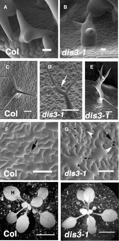
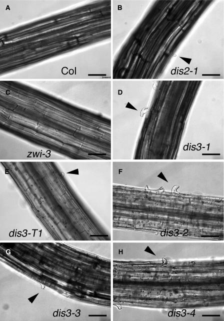
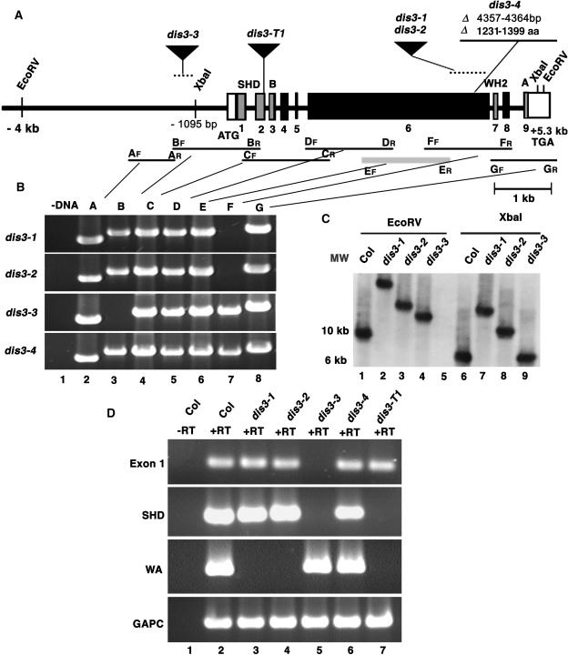
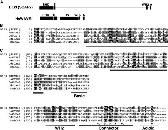
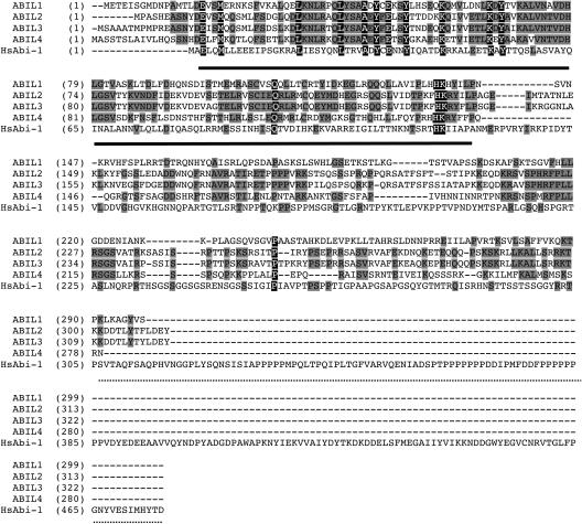
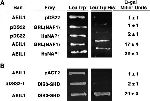
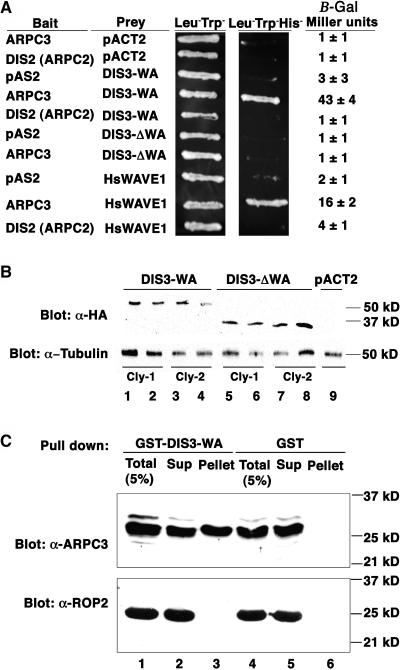
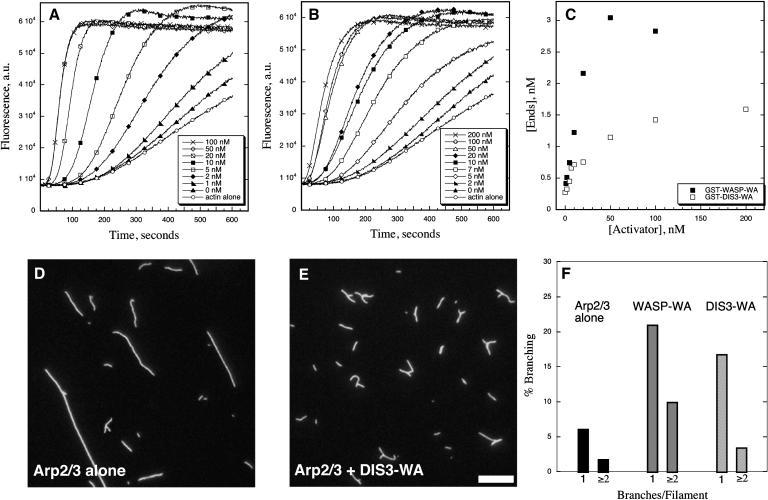
In a plant cell, a subset of actin filaments function as a scaffold that positions the endomembrane system and acts as a substrate on which organelle motility occurs. Other actin filament arrays appear to be more dynamic and reorganize in response to growth signals and external cues. The distorted group of trichome morphology mutants provides powerful genetic tools to study the control of actin filament nucleation in the context of morphogenesis. In this article, we report that DISTORTED3 (DIS3) encodes a plant-specific SCAR/WAVE homolog. Null alleles of DIS3, like those of other Arabidopsis thaliana WAVE and Actin-Related Protein (ARP) 2/3 subunit genes, cause trichome distortion, defects in cell-cell adhesion, and reduced hypocotyl growth in etiolated seedlings. DIS3 efficiently activates the actin filament nucleation and branching activity of vertebrate Arp2/3 and functions within a WAVE-ARP2/3 pathway in vivo. DIS3 may assemble into a WAVE complex via a physical interaction with a highly diverged Arabidopsis Abi-1-like bridging protein. These results demonstrate the utility of the Arabidopsis trichome system to understand how the WAVE and ARP2/3 complexes translate signaling inputs into a coordinated morphogenetic response.


Figures

Figure 1.
Figure 1.
Mutation of DISTORTED3 Causes Trichome Distortion and Defective Cell–Cell Adhesion in the Cotyledon Epidermis. (A) Scanning electron micrograph of a wild-type stage 4 trichome that contains young branches with a blunt tip morphology. (B) A representative stage 4 dis3-1 trichome with a blunt tip morphology and a swollen stalk. (C) Mature wild-type (Columbia-0 [Col-0]) trichome displaying three highly elongated and tapered branches. (D) An example of the mild dis3-1 mature trichome phenotype. Trichomes in this class display an abnormally elongated interbranch zone (arrow) and mild branch swelling. (E) An example of the severe dis3-1 mature trichome phenotype. Branch lengths are reduced and slightly swollen. The interbranch zone is abnormally elongated and swollen. (F) Upper surface of a representative 12-DAG wild-type cotyledon. Pavement cells are highly lobed. Image was taken from the most highly expanded cells in the apical third of the cotyledon. (G) Upper surface of representative 12-DAG dis3-1 cotyledon pavement cells. Image was taken from the most highly expanded cells in the apical third of the cotyledon. (H) Low magnification image of a representative 14-DAG wild-type seedling. (I) Low magnification image of a representative 14-DAG dis3-1 seedling. The white arrowheads label the gaps between adjacent pavement cells, black arrows label stomatal pores, and white arrows label the interbranch zone of a dis3 trichome. Bars = 10 μm in (A) and (B), 100 μm in (C) to (H), and 1 cm in (I) and (J).
Figure 2.
Figure 2.
Cell–Cell Adhesion Defects in Etiolated Hypocotyls of dis3 Alleles. (A) Wild-type hypocotyls with adherent epidermal cells. (B) Epidermal cell–cell adhesion defects in dis2-1 (arpc2). (C) zwi-3 hypocotyls with adherent epidermal cells. (D) Epidermal cell–cell adhesion defects in dis3-1. (E) Epidermal cell–cell adhesion defects in dis3-T1. (F) Epidermal cell–cell adhesion defects in dis3-2. (G) Epidermal cell–cell adhesion defects in dis3-3. (H) Epidermal cell–cell adhesion defects in dis3-4. Differential interference contrast images of whole-mounted, 7-DAG, dark-grown seedlings. Arrowheads indicate the location of cell–cell adhesion defects. Bar = 100 μm in all panels.
Figure 3.
Figure 3.
Physical Map and Molecular Characterization of DIS3/SCAR2 Mutant Alleles. (A) The physical structure of the DIS3/SCAR2 gene. The transcribed region of DIS3/SCAR2 is labeled with boxes (exons) and lines (introns). The DIS3/SCAR2 exons that encode conserved domains or motifs are gray and are labeled with text: SHD, SCAR homology domain; B, basic region; WH2, G-actin binding region; A, acidic region. The location and nature of dis3-1, dis3-2, dis3-3, dis3-4, and dis3-T1 alleles are defined. Nucleotides are numbered relative to the first nucleotide of the start codon defined as +1. aa, amino acids. (B) The dis3-1 through dis3-4 alleles disrupt the DIS3/SCAR2 gene. Genomic DNA was isolated from dis3-1, dis3-2, dis3-3, and dis3-4 alleles, and overlapping pairs of PCR primers that spanned the DIS3/SCAR2 locus were used to locate the affected regions of the gene. The pairs of PCR primers are named according to their order along the gene and their direction, either forward (F) or reverse (R). Lane 1 includes a representative no template DNA control PCR reaction, and lanes 2 to 8 contain the PCR products obtained using PCR primer pairs A through G, respectively. The location of each primer pair relative to the DIS3/SCAR2 gene is indicated in (A). (C) The dis3-1, dis3-2, and dis3-3 alleles contain DNA insertions. DNA gel blot analysis was conducted on genomic DNA that was isolated from Col, dis3-1, dis3-2, and dis3-3 seedlings. Genomic DNA was digested with EcoRV (lanes 1 to 4) and XbaI (lanes 6 to 9). The probe was constructed using PCR product E shown in (A). MW, DNA size standards. (D) The dis3 alleles that harbor DNA insertions affect the DIS3/SCAR2 transcript. RNA was isolated from Col and dis3 mutant seedlings and subjected to RT-PCR analyses using primer pairs located within different regions of the DIS3/SCAR2 gene, including exon 1, primers that flank the encoded SCAR homology domain, and primers that flank the WA domain. Glyceraldehyde-3-phosphate dehydrogenase C subunit (GAPC) was used as a positive control. Lane 1, a representative no reverse transcriptase (−RT) control; lanes 2 to 7, plus reverse transcriptase (+RT) for Col, dis3-1, dis3-2, dis3-3, dis3-4, and dis3-T1, respectively.
Figure 4.
Figure 4.
DIS3/SCAR2 Is a SCAR Homology and WA Domain–Containing Protein. (A) The domain organization and relative sizes of the DIS3/SCAR2 and human WAVE1. The abbreviations of the domains are as in Figure 3. Pr, Pro-rich region of human WAVE. (B) The SCAR homology domains of DIS3/SCAR2 contain blocks of highly conserved amino acid sequences. Amino acid sequences of the SCAR homology domain of DIS3/SCAR2 and other WAVE/SCAR family proteins were aligned using ClustalW. The bold black line and the double lines mark two highly conserved blocks toward the N- and C-terminal ends of the SCAR homology domains, respectively. A region enriched in basic amino acids present in all SCAR/WAVE proteins is underlined and labeled. (C) DIS3/SCAR2A contains a C-terminal WA domain. Amino acid sequences of DIS3/SCAR2 and other WAVE/SCAR family proteins were aligned using ClustalW. According to published alignments and functional studies of the WA domain (Panchal et al., 2003), the alignments were anchored using a conserved Arg (marked B below the alignment) and Trp (marked W in the alignment) residues, and conserved hydrophobic amino acids (marked Φ) in the connector region are labeled (Panchal et al., 2003). The WH2 G-actin binding region and acidic regions are labeled. The GenBank accession numbers used for alignments are AY817016 (DIS3/SCAR2), AAD33052 (human WAVE1), NP_493028 (C. elegans WVE-1), AAD29083 (D. discoideum Scar1), and AAF74194 (D. melanogaster SCAR).
Figure 5.
Figure 5.
SCAR Genes Are Expressed throughout Plant Development. Total RNA isolated from the organs listed at the top of the figure was analyzed by RT-PCR using gene-specific primers for SCAR1, SCAR2, SCAR3, and SCAR4. PIR (SRA-1) expression was analyzed to provide an example for a WAVE complex gene. DIS2 (ARPC2) and ARPC4 were used as examples of ARP2/3 subunit genes. For each primer pair the PCR cycle number was optimized to reveal relative differences in expression between organs and were SCAR1 (35 cycles), SCAR2 (35 cycles), SCAR3 (35 cycles), SCAR4 (35 cycles), ABIL1 (25 cycles), PIR (SRA-1) (22 cycles), DIS2 (ARPC2) (20 cycles), ARPC4 (21 cycles), and GAPC (glyceraldehyde-3-phosphate dehydrogenase C subunit) (20 cycles). Lane 1, no reverse transcriptase (−RT) control; lanes 2 to 10, various RNA samples subjected to RT treatment. Control experiments that lacked RT were conducted on all RNA samples, but only the seedling experiment is shown.
Figure 6.
Figure 6.
A Subpopulation of dis3 Trichomes Fails to Generate and Maintain a Normal Population of Cytoplasmic Actin Bundles. The actin cytoskeletons of whole-mounted fixed cells and living stage 3/4 trichomes were detected using Alexa-488 phalloidin ([A] to [H]) and GFP:ABD2 ([I] to [N]) and visualized using confocal microscopy. For each actin probe, the panels on the left are maximum projections of an entire cell, and those on the right include the midplanes of longitudinal optical sections through the branch that is boxed in the maximum projection. Arrows in (G) and (H) indicate actin bundles that have a full or partial cortical localization. Bar =10 μm in all panels. (A) and (B) F-actin organization in a wild-type stage 3/4 trichome visualized using fluorescent phalloidin. (C) and (D) A dis3-1 stage 3/4 trichome displaying a wild type–like parallel actin bundle organization. (E) and (F) A dis3-1 trichome displaying disorganized actin filaments and/or bundles. (G) and (H) A dis2-1 (arpc2) trichome displaying mislocalized cytoplasmic actin filaments. (I) and (J) Actin organization of a wild-type stage 3/4 trichome visualized using GFP:ABD2. (K) and (L) A dis3-1 GFP:ABD2-expressing plant containing a stage 3/4 trichome displaying a wild type–like parallel actin bundle organization. (M) and (N) A dis3-1 GFP:ABD2-expressing plant—a representative example of a stage 3/4 trichome lacking a population of loosely aligned cytoplasmic bundles.
Figure 7.
Figure 7.
Elongating Stage 5 dis3-1 Trichomes Have Defects in Actin Filament Organization and Vacuole Positioning. Whole-mounted fixed trichomes belonging to this stage were identified based on a pointed tip morphology, the absence of papillae, a cell height of at least 40 μm, and a branch length between 30 and 75 μm. Left panels are maximum projections of the entire branch, and those on the right include longitudinal optical sections through the middle of the branch within the boxed region. (A) and (B) Actin organization in a wild-type trichome. (C) and (D) A dis3-1 stage 5 trichome displaying aligned core actin filaments. (E) and (F) A dis3-1 trichome in which the core cytoplasm lacks actin bundles and is occupied by the central vacuole. Bar = 10 μm in (A) to (F). (G) Quantitation of the relative amounts of cortical and core cytoplasmic actin in Alexa 488 phalloidin–labeled samples. Total integrated intensity of core: total actin signal was measured in several image planes centered on the branch midpoint. Intensity ratios for Col and dis3 branches were measured in stage 3/4 and stage 4/5 trichomes. Mean value of each measured branch is plotted and the grand mean ± sd for each genotype is listed in the plot. (H) Same as in (G) but calculated using GFP:ABD as a probe. Asterisks indicate significant difference compared with the wild type according to a Student's t test (P value < 0.05).
Figure 8.
Figure 8.
Microtubule Organization in Wild-Type and dis3 Trichomes at Different Stages. Microtubules were visualized in living cells using the GFP:MBD probe. All images are maximum projections of confocal images. Bar = 10 μm in all panels. (A) Transversely aligned cortical microtubules in a stage 4 wild-type trichome. (B) Oblique alignment of microtubules in a wild-type stage 5 trichome. (C) Microtubule organization in a dis3-1 stage 4 trichome with a normal branch morphology. (D) Microtubule organization in a dis3-1 stage 4 trichome that displays a reduced branch length (arrow) and branch swelling (arrowhead). (E) Microtubule arrangements in stage 5 dis3-1 trichomes. Projections of two cells on the same leaf that have either no obvious microtubule defect (arrow) or a swollen morphology and randomly organized microtubules (arrowhead).
Figure 9.
Figure 9.
Amino Acid Sequence Alignment of the Four Arabidopsis Proteins that Share Limited Amino Acid Identity with the N Terminus of Human Abi-1. The N-terminal region of conserved amino acid identity between the Arabidopsis proteins and human Abi-1 is underlined. The SH3 domain of Abi-1 is labeled with a dashed line. GenBank accession numbers are listed in Methods.
Figure 10.
Figure 10.
An Arabidopsis Abi-1–Like Protein May Mediate the Assembly of DIS3/SCAR2 into a WAVE Complex. (A) ABIL1 interacts with GRL (NAP1) and human NAP1 in the yeast two-hybrid assay. The bait and prey plasmids that were cotransformed into yeast are defined to the left of the corresponding yeast patches, and β-galactosidase assay results are shown to the right. (B) The SHD domain of DIS3/SCAR2 interacts with ABIL1 in the yeast two-hybrid assay. The bait and prey plasmids that were cotransformed into yeast are defined to the left of the corresponding yeast patches, and β-galactosidase assay results are shown to the right.
Figure 11.
Figure 11.
The WA Domain of DIS3/SCAR2 Is Required for Direct Binding to ARPC3. (A) DIS3/SCAR2 and human WAVE1 interact with ARPC3 in a yeast two-hybrid assay. The bait and prey plasmids that were cotransformed into yeast are defined to the left of the corresponding yeast patches and β-galactosidase assay results to the right. DIS3-WA contains amino acids 1032 to 1399; DIS3-ΔWA contains amino acids 1032 to 1238; ARPC3, DIS2 (ARPC2), and HSWAVE1 are full-length proteins. (B) The DIS3-ΔWA and DIS3-WA two-hybrid constructs are expressed at comparable levels in yeast. Top, lanes 1 to 9, duplicate yeast protein soluble fractions probed with anti-HA antibody. Bottom, same blot probed with control anti-tubulin antibody. Lanes 1-4, duplicate protein samples extracted from two individual colonies (cly-1 and cly-2) of the yeast strain expressing HA-tagged DIS3-WA; lanes 5 to 8, same as 1 to 4, but expressing HA-tagged DIS3-ΔWA; lane 9, yeast extracts from the strain harboring the empty vector pACT2. All the strains used also contained the ARPC3 bait construct. (C) Direct interaction of DIS3/SCAR2 and ARPC3 in a GST pull-down assay. Top, lanes 1 to 6, fractions probed with an anti-ARPC3 antibody; bottom, fractions probed with an anti-ROP2 antibody. Lane 1, 5% of total binding reaction; lane 2, unbound; lane 3, GST-DIS3-WA–associated fraction; lane 4, 5% of total binding reaction with GST alone control beads; lane 5, unbound; lane 6, GST bead-associated fraction.
Figure 12.
Figure 12.
The WA Domain of DIS3/SCAR2 Efficiently Activates Vertebrate Arp2/3. (A) to (C) Actin (4 μM) (5% pyrene labeled) with or without 50 nM Arp2/3 and various amounts of activators was allowed to polymerize and the kinetics of assembly monitored by fluorimetry. Pyrene fluorescence (a.u., arbitrary units), which increases when actin assembles into filaments, is plotted versus time after addition of G-actin to initiate polymerization. Nucleation activity is evident as a decrease in the lag period required before actin assembly ensues and an increase in the initial rate of polymerization. (A) Polymerization of actin in the presence of 50 nM Arp2/3 and a GST fusion protein with the WA domain of human N-WASP (GST-WASP-WA) at various concentrations: 0, 1, 2, 5, 10, 20, 50, and 100 nM (see legend). (B) Similar experiments performed in the presence of the WA domain from DIS3 (GST-DIS3-WA) at various concentrations: 0, 2, 5, 7, 10, 20, 50, 100, and 200 nM (see legend). DIS3-WA was comprised of amino acids 1084 to 1399 of DIS3/SCAR2. (C) Dependence of the concentration of apparent ends ([Ends]) on the concentration of WASP-WA (closed squares) and DIS3-WA (open squares) calculated from the rate of polymerization at the time where 50% (2.0 μM) of the actin was polymerized. These values were calculated from the data shown in (A) and (B) using the equation described in Methods. (D) to (F) Activators of Arp2/3 were also analyzed by fluorescence microscopy for the ability to stimulate branch formation. Conditions: 4 μM G-actin was polymerized in the presence of 50 nM Arp2/3 with or without activators. Equimolar rhodamine-phalloidin was added to the reactions to label filaments, and reactions were diluted into fluorescence buffer after 30 min. (D) Arp2/3 alone (50 nM), no activator (E) Arp2/3 (50 nM) + 100 nM GST-DIS3-WA. Bar = 2.5 μm. (F) The bar graph shows quantification of branch frequency counted from at least five different micrographs and >300 filaments for each treatment. Percentage of branching was binned into three categories: no branches, one branch, and two or more branches. Given on the graph are the latter two subcategories.

References

    1. Alonso, J.M., et al. (2003). Genome-wide insertional mutagenesis of Arabidopsis thaliana. Science 301, 653–657. - PubMed
    1. Ausubel, F.M., Brent, R., Kingston, R.E., Moore, D.D., Seidman, J.G., Smith, J.A., and Struhl, K. (1994). Current protocols in molecular biology. (New York: John Wiley & Sons).
    1. Basu, D., El-Assal, S.E., Le, J., Mallery, E.L., and Szymanski, D.B. (2004). Interchangeable functions of Arabidopsis PIROGI and the human WAVE complex subunit SRA-1 during leaf epidermal morphogenesis. Development 131, 4345–4355. - PubMed
    1. Ben-Yaacov, S., Le Borgne, R., Abramson, I., Schweisguth, F., and Schejter, E. (2001). Wasp, the Drosophila Wiskott-Aldrich syndrome gene homologue, is required for cell fate decisions mediated by notch signaling. J. Cell Biol. 152, 1–13. - PMC - PubMed
    1. Blagg, S.L., and Insall, R.H. (2004). Solving the WAVE function. Nat. Cell Biol. 6, 279–281. - PubMed

NOTE ADDED IN PROOF

    1. While this work was under review, Frank et al. (2004) reported that plant SCAR proteins bind to BRK1 (HSPC300) and can activate vertebrate Arp2/3.
    1. Frank, M., Egile, C., Dyachok, J., Djakovic, S., Nolasco, M., Li, R., and Smith, L.G. (2004). Activation of Arp2/3 complex-dependent actin polymerization by plant proteins distantly related to Scar/WAVE. Proc. Natl. Acad. Sci. USA 101, 16379–16384. - PMC - PubMed

Publication types

MeSH terms

Associated data