. 2005 Apr;16(4):1673-83.
doi: 10.1091/mbc.e04-10-0873. Epub 2005 Jan 19.

Yos1p is a novel subunit of the Yip1p-Yif1p complex and is required for transport between the endoplasmic reticulum and the Golgi complex

Affiliations

Yos1p is a novel subunit of the Yip1p-Yif1p complex and is required for transport between the endoplasmic reticulum and the Golgi complex

Matthew Heidtman et al. Mol Biol Cell. 2005 Apr.

Abstract

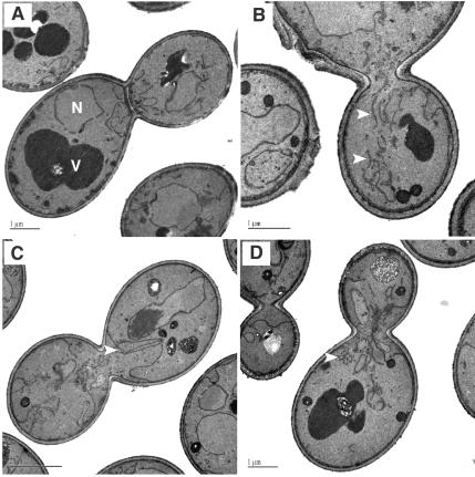
Yeast Yip1p is a member of a conserved family of transmembrane proteins that interact with Rab GTPases. Previous studies also have indicated a role for Yip1p in the biogenesis of endoplasmic reticulum (ER)-derived COPII transport vesicles. In this report, we describe the identification and characterization of the uncharacterized open reading frame YER074W-A as a novel multicopy suppressor of the thermosensitive yip1-4 strain. We have termed this gene Yip One Suppressor 1 (YOS1). Yos1p is essential for growth and for function of the secretory pathway; depletion or inactivation of Yos1p blocks transport between the ER and the Golgi complex. YOS1 encodes an integral membrane protein of 87 amino acids that is conserved in eukaryotes. Yos1p localizes to ER and Golgi membranes and is efficiently packaged into ER-derived COPII transport vesicles. Yos1p associates with Yip1p and Yif1p, indicating Yos1p is a novel subunit of the Yip1p-Yif1p complex.


Figures

Figure 1.
Figure 1.
YOS1 is a multicopy suppressor of yip1-4. (A) Strains were grown to saturation in minimal media lacking uracil to maintain selection of plasmids, adjusted to an OD600 of 1.0, and 5 μl of a 10-fold dilution series was spotted onto YPD plates. Plates were then incubated at 25 or 34°C for 48 h. (B) Multicopy YOS1 partially rescues the budding defect in the yip1-4 strain. Wash semi-intact cell membranes from different strains containing [35S]gpαf were incubated with COPII proteins and an energy regeneration system to measure vesicle budding. After 30 min at 23°C, freely diffusible vesicles containing [35S]gpαf were separated from semi-intact cell membranes by centrifugation at 18,000 × g, and [35S]gpαf was quantified by concanavalin A precipitation.
Figure 2.
Figure 2.
Sequence comparison of Yos1p from different species. The amino acid sequence of Yos1p from S. cerevisiae is aligned with those of related proteins from Schizosaccharomyces pombe SPAC19A8.09, Caenorhabditis elegans W09G3.8, Drosophila melanogaster CG32069, and Homo sapiens HSPC039. Alignments were generated using the ClustalW program. Invariant residues are shaded in black, whereas regions in which conserved substitutions have been observed are shaded in gray. Lines indicate predicted transmembrane segments.
Figure 3.
Figure 3.
YOS1 is an essential gene and is required for protein transport between the ER and the Golgi complex. (A) A YOS1/yos1Δ strain was transformed with empty vector or a centromere-based plasmid bearing wild-type YOS1. Transformants were sporulated, and asci were dissected on YPD plates at 25°C. The YOS1/yos1Δ strain transformed with empty vector (pRS316) displayed a 2+:2 segregation pattern, indicating YOS1 is an essential gene (left). Inviability could be rescued in certain cases by the presence of pRS316/YOS1 (right), demonstrating that the 2+:2 segregation pattern is a specific consequence of the disruption of YOS1. (B) Depletion of Yos1p causes a block in protein transport between the ER and the Golgi complex. A GAL1-YOS1 strain and an isogenic wild-type strain were grown overnight in media containing 2% galactose/2% glucose. Cells were then washed and back diluted to an OD600 of 0.1 in media containing 2% glucose to induce transcriptional silencing of the GAL1 promoter-controlled YOS1 gene and allowed to grow at 30°C. Equivalent OD600 units were harvested from each strain at various time points after shift to glucose. Membrane fractions were prepared and analyzed by immunoblot for the secretory proteins CPY and Gas1p. Sec61p was immunoblotted as a loading control, whereas immunoblot of Yos1p is a control to show efficient depletion of Yos1p upon GAL1 repression.
Figure 4.
Figure 4.
Yos1p is an integral membrane protein. (A) Hydrophobicity plot of the Yos1p amino acid sequence as described previously (Kyte and Doolittle, 1982). (B) Microsomes prepared from wild-type cells (FY834) were treated with buffer (B88), 0.1 M Na2CO3 (pH 11), or buffer containing 1% Triton X-100 (1% TX) and centrifuged at 100,000 × g. Totals before centrifugation (T), supernatant (S), and pellet fractions (P) were analyzed on a 15% polyacrylamide gel and immunoblotted for Sec23p (peripheral membrane protein), Yip1p (integral membrane protein), and Yos1p.
Figure 5.
Figure 5.
Yos1p localizes to the early secretory pathway. (A) Yos1p fractionates with ER and Golgi membranes. A whole cell lysate from wild-type cells was separated on a sucrose density gradient (20–60%), and fractions were collected from the top. Relative levels of Sec61p (ER marker), Och1p (Golgi marker), Yos1p, and Yip1p were quantified by densitometry of immunoblots. (B) Yos1p is packaged into COPII-coated vesicles. COPII-dependent budding reactions were performed from wild-type (FY834) microsomes. One-tenth of a total reaction (T) and budded vesicles isolated after a mock incubation (–) or after incubation with COPII proteins (+) were resolved on a 15% polyacrylamide gel. Sec61p (ER-resident protein), Sec22p (SNARE protein), Yip1p, and Yos1p were detected by immunoblot.
Figure 6.
Figure 6.
Yos1p forms a complex with Yip1p and Yif1p. Solubilized proteins were bound to anti-HA or anti-Yos1p antibodies coupled to protein A beads (IP) or beads alone (–) as described in Materials and Methods. (A) Immunoblots of native anti-HA immunoprecipitations from YIF1–3×HA and untagged (WT) solubilized microsomes. Total lanes (T) and solubilized (S) lanes represent 1.5% of the starting material. Note specific coimmunoprecipitation of Yip1p and Yos1p with Yif1p-3×HA. (B) Immunoblots of native anti-Yos1p immunoprecipitations from YIF1-3×HA microsomes. T and S lanes represent 1.5% of the starting material. Note the specific coimmunoprecipitation of Yip1p and Yif1p-3×HA with Yos1p. Asterisk indicates antibody heavy chain cross-reactivity with secondary antibodies.
Figure 7.
Figure 7.
Yos1p interaction with Yip1p is disrupted in the yip1-4 strain. Solubilized proteins were bound to anti-Yos1p antibodies coupled to protein A beads (IP) or beads alone (–) as described in Materials and Methods. Total lanes (T) and Solubilized (S) lanes represent 3% of the starting material. Note the specific coimmunoprecipitation of Yip1p with Yos1p from wild-type membranes, whereas this interaction is significantly reduced in the yip1-4 strain.
Figure 8.
Figure 8.
Mutant yos1-1 cells display transport defects in vivo and in vitro. (A) Pulse-chase analysis of CPY maturation in wild-type and yos1-1 strains. Wild-type (CBY1766) and yos1-1 (CBY1822) strains were pulsed for 7 min with 35S-labeled methionine and then chased for 30 min. Labeled CPY was immunoprecipitated from cell extracts, resolved on an 8% acrylamide gel, and visualized by autoradiography. (B) Washed semi-intact cells containing [35S]gpαf were prepared from wild-type (CBY1766) and yos1-1 (CBY1822) strains. Semi-intact cells were incubated with COPII proteins, Uso1p, LMA1, and an ATP regeneration system. After 80 min at 25°C, the amount of Golgi-modified [35S]gpαf was measured to determine transport efficiency. (C) Semi-intact cells from wild-type or mutant strains were prepared as in A and incubated with COPII or COPII plus Uso1p to measure vesicle budding and tethering. After 30 min at 25°C, freely diffusible vesicles containing [35S]gpαf were separated from semi-intact cell membranes by centrifugation at 18,000 × g and [35S]gpαf quantified by concanavalin A precipitation.
Figure 9.
Figure 9.
Yip1p interaction with Yos1p is disrupted in the yos1-1 strain. Solubilized proteins were bound to anti-Yip1p antibodies coupled to protein A beads (IP) or beads alone (–) as described in Materials and Methods. Total lanes (T) and Solubilized (S) lanes represent 3% of the starting material. Note the specific coimmunoprecipitation of Yos1p with Yip1p from wild-type membranes, whereas this interaction is absent in the yos1-1 strain. Asterisks indicate antibody heavy chain cross-reactivity with secondary antibodies.
Figure 10.
Figure 10.
Mutant yos1-1 cells display morphological phenotypes characteristic of a block in the early secretory pathway. Wild-type (A) and mutant cells (B–D) were shifted to 37°C for 60 min and then fixed and prepared for EM as described in Materials and Methods. Representative thin sections are shown for each condition. The white lettering in A denotes the nucleus (N) and vacuole (V). The white arrows in B and C highlight dislocated tubular ER elements, whereas the white arrow in D highlights a cluster of vesicular structures. Bars, 1 μm (A, B, and D) and 2 μm (C).

Similar articles

Cited by

References

    1. Ausubel, R. M., Brent, R., Kingston, R. E., Moore, D. D., Seidman, J. G., Smith, J. A., and Struhl, K. (1987). Current Protocols in Molecular Biology, New York, Greene Publishing Associates and Wiley-Interscience.
    1. Baker, D., Hicke, L., Rexach, M., Schleyer, M., and Schekman, R. (1988). Reconstitution of SEC gene product-dependent intercompartmental protein transport. Cell 54, 335–344. - PubMed
    1. Barlowe, C. (1997). Coupled ER to Golgi transport reconstituted with purified cytosolic proteins. J. Cell Biol. 139, 1097–1108. - PMC - PubMed
    1. Barrowman, J., Wang, W., Zhang, Y., and Ferro-Novick, S. (2003). The Yip1p·Yif1p complex is required for the fusion competence of endoplasmic reticulum-derived vesicles. J. Biol. Chem. 278, 19878–19884. - PubMed
    1. Becker, J., Tan, T. J., Trepte, H. H., and Gallwitz, D. (1991). Mutational analysis of the putative effector domain of the GTP-binding protein Ypt1p in yeast suggests specific regulation by a novel GAP activity. EMBO J. 10, 785–792. - PMC - PubMed

Publication types

MeSH terms

Substances