. 2005 Feb;187(3):829-39.
doi: 10.1128/JB.187.3.829-839.2005.

Disparate subcellular localization patterns of Pseudomonas aeruginosa Type IV pilus ATPases involved in twitching motility

Affiliations

Disparate subcellular localization patterns of Pseudomonas aeruginosa Type IV pilus ATPases involved in twitching motility

Poney Chiang et al. J Bacteriol. 2005 Feb.

Abstract

The opportunistic pathogen Pseudomonas aeruginosa expresses polar type IV pili (TFP), which are responsible for adhesion to various materials and twitching motility on surfaces. Twitching occurs by alternate extension and retraction of TFP, which arise from assembly and disassembly of pilin subunits at the base of the pilus. The ATPase PilB promotes pilin assembly, while the ATPase PilT or PilU or both promote pilin dissociation. Fluorescent fusions to two of the three ATPases (PilT and PilU) were functional, as shown by complementation of the corresponding mutants. PilB and PilT fusions localized to both poles, while PilU fusions localized only to the piliated pole. To identify the portion of the ATPases required for localization, sequential C-terminal deletions of PilT and PilU were generated. The conserved His and Walker B boxes were dispensable for polar localization but were required for twitching motility, showing that localization and function could be uncoupled. Truncated fusions that retained polar localization maintained their distinctive distribution patterns. To dissect the cellular factors involved in establishing polarity, fusion protein localization was monitored with a panel of TFP mutants. The localization of yellow fluorescent protein (YFP)-PilT and YFP-PilU was independent of the subunit PilA, other TFP ATPases, and TFP-associated proteins previously shown to be associated with the membrane or exhibiting polar localization. In contrast, YFP-PilB exhibited diffuse cytoplasmic localization in a pilC mutant, suggesting that PilC is required for polar localization of PilB. Finally, localization studies performed with fluorescent ATPase chimeras of PilT and PilU demonstrated that information responsible for the characteristic localization patterns of the ATPases likely resides in their N termini.


Figures

FIG. 1.
FIG. 1.
Complementation of twitching motility by fluorescent fusion constructs in pilB, pilT, and pilU mutants. Thin (3-mm) 1% LB agar plates were stab inoculated with a toothpick and incubated at 37°C for 48 h (47). Twitching motility was visualized as a halo surrounding the point of inoculation, between the agar and the plastic plate. Both twitching and nontwitching strains formed colonies on the agar surface. A, pilT mutant carrying pilT-cfp; B, pilT mutant carrying cfp-pilT; C, wild-type strain PAK carrying yfp-pilT; D, wild-type strain PAK; E, pilT mutant; F, pilT mutant carrying yfp-pilT; G, pilT mutant carrying pilT; H, pilB mutant; I, pilB mutant carrying yfp-pilB; J, pilB mutant carrying pilB; K, wild-type strain PAK; L, pilU mutant; M, pilU mutant carrying yfp-pilU; N, pilU mutant carrying pilU. Scale bar, 10 mm.
FIG. 2.
FIG. 2.
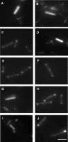
P. aeruginosa ATPase mutants carrying fluorescent protein fusions to PilB, PilT, or PilU. (A) pilT mutant carrying cfp. (B) pilT mutant carrying pilT-cfp. (C) pilT mutant carrying cfp-pilT. (D) pilT mutant carrying yfp. (E) pilT mutant carrying yfp-pilT. (F) Same as panel E with punctuate fluorescence at the division septum. (G) pilB mutant carrying yfp. (H) pilB mutant carrying yfp-pilB. (I) pilU mutant carrying yfp. (J) pilU mutant carrying yfp-pilU. Scale bar, 2 μm.
FIG. 3.
FIG. 3.
Fluorescent time-lapse images of free-swimming pilU mutant carrying yfp-pilU. A single mutant pilU cell carrying yfp-pilU swam from left to right via flagellum-mediated motility. The image is a composite of single time-lapse images taken at 1-s intervals. The arrows point to the trailing piliated or flagellated end of the cell at 0 and 4 s. Scale bar, 5 μm.
FIG. 4.
FIG. 4.
Localization of YFP-PilB in a pilC mutant. (A) Wild-type strain PAK carrying yfp-pilB. (B) pilB mutant carrying yfp-pilB. (C) pilC mutant carrying yfp-pilB. Scale bars, 2 μm.
FIG. 5.
FIG. 5.
Schematic representation of truncated fusion proteins used in this study. The full-length fusion with characteristic AAA+ protein motifs is shown at the top. The positions of Walker box A (A), the Asp box (Asp), Walker box B (B), and the His box (His) are indicated. YFP-PilU deletions and YFP-PilT deletions are also shown. The localization pattern of each deletion construct is indicated on the right. None of the deletions could complement the corresponding mutants.
FIG. 6.
FIG. 6.
Predicted structures of PilT and PilU. The structures of PilT and PilU were predicted based on the recently solved EpsE (V. cholerae T2S ATPase) structure by using the 3D-PSSM fold recognition server. A significant match in the structure of PilT and PilU with EpsE was obtained (PSSM e-value, 1.59e-05; 95% certainty). All three ATPases have globular N- and C-terminal domains bridged by a central linker region. (A) Structure of EpsE. (B) Predicted structure of PilT. (C) Predicted structure of PilU.
FIG. 7.
FIG. 7.
Localization of PilT and PilU chimeras. (A) pilT mutant carrying yfp-N′pilT-C′pilU. (B) pilU mutant carrying yfp-N′pilT-C′pilU. (C) pilT mutant carrying yfp-N′pilU-C′pilT. (D) pilU mutant carrying yfp-N′pilU-C′pilT. Scale bars, 2 μm.
FIG. 8.
FIG. 8.
LB agar plate morphology of pilT and pilU cultures. Cells were streaked on LB agar and incubated at 37°C for 24 h. Regions A, B, and C contained the PAK strain of P. aeruginosa and derivatives of this strain, while regions D, E, and F contained the mPAO1 strain and derivatives of this strain. (A and D) pilU mutants. (B and E) Wild-type strain. (C and F) pilT mutants.

Similar articles

Cited by

References

    1. Ball, G., V. Chapon-Herve, S. Bleves, G. Michel, and M. Bally. 1999. Assembly of XcpR in the cytoplasmic membrane is required for extracellular protein secretion in Pseudomonas aeruginosa. J. Bacteriol. 181:382-388. - PMC - PubMed
    1. Bhattacharjee, M. K., S. C. Kachlany, D. H. Fine, and D. H. Figurski. 2001. Nonspecific adherence and fibril biogenesis by Actinobacillus actinomycetemcomitans: TadA protein is an ATPase. J. Bacteriol. 183:5927-5936. - PMC - PubMed
    1. Boyd, J. M. 2000. Localization of the histidine kinase PilS to the poles of Pseudomonas aeruginosa and identification of a localization domain. Mol. Microbiol. 36:153-162. - PubMed
    1. Boyd, J. M., and S. Lory. 1996. Dual function of PilS during transcriptional activation of the Pseudomonas aeruginosa pilin subunit gene. J. Bacteriol. 178:831-839. - PMC - PubMed
    1. Bradley, D. E. 1980. A function of Pseudomonas aeruginosa PAO polar pili: twitching motility. Can. J. Microbiol. 26:146-154. - PubMed

Publication types

MeSH terms

LinkOut - more resources