. 2005 Feb;187(3):1022-35.
doi: 10.1128/JB.187.3.1022-1035.2005.

The Enterococcus faecalis sigV protein is an extracytoplasmic function sigma factor contributing to survival following heat, acid, and ethanol treatments

Affiliations

The Enterococcus faecalis sigV protein is an extracytoplasmic function sigma factor contributing to survival following heat, acid, and ethanol treatments

Abdellah Benachour et al. J Bacteriol. 2005 Feb.

Abstract

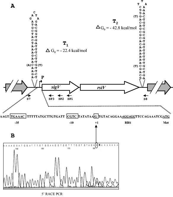
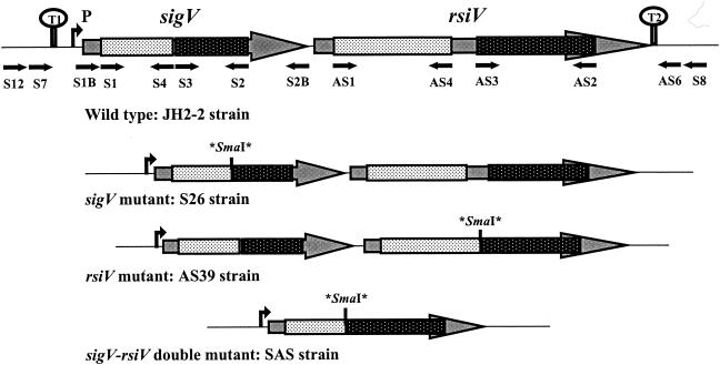
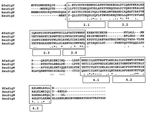
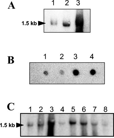
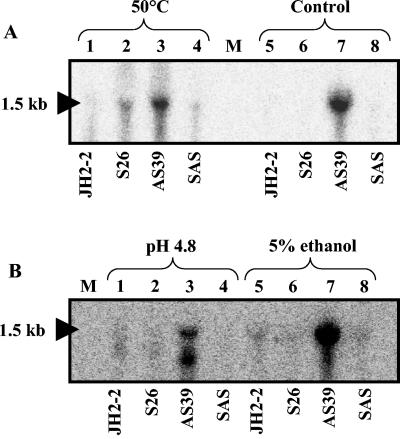
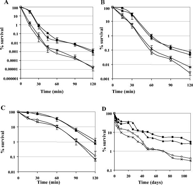
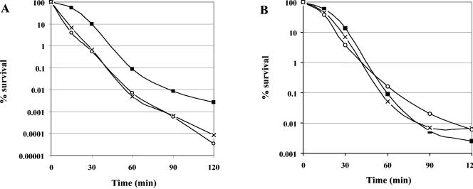
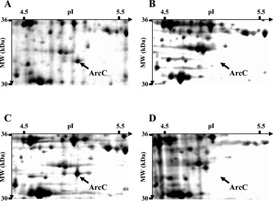
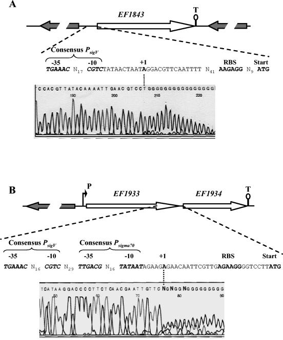
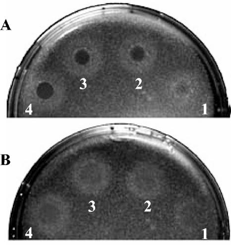
Analysis of the genome sequence of Enterococcus faecalis allowed the identification of two genes whose protein products showed 33 and 34% identity with those of sigV and yrhM of Bacillus subtilis, respectively. These genes, named sigV and rsiV, are predicted to encode members of the extracytoplasmic function subfamily of eubacterial RNA polymerase sigma and anti-sigma factors, respectively. This group of sigma factors has been shown to regulate gene expression in response to stress conditions. sigV and rsiV were shown to be under the control of the same promoter. The transcriptional start site was determined, and the 1.5-kb mRNA transcript was shown to be overexpressed under glucose and complete starvation, as well as under physicochemical treatments. Three mutants, affected in sigV, rsiV, and both genes, were constructed by double-crossover recombination within the genome of E. faecalis strain JH2-2. Compared with the wild type and the rsiV mutant, the sigV mutants were more susceptible to heat shock, acid, and ethanol treatments and displayed decreased survival during long-term starvation. A nisin-inducible sigV gene construction used in complementation assays restored the wild phenotype of the sigV mutants, confirming the involvement of SigV in the heat shock, ethanol, and acid stress responses. Northern blot analysis carried out with the three mutant strains revealed the inhibition of sigV expression by the related anti-sigma factor gene rsiV. In addition, putative candidates of the sigV regulon determined by computer search for the sigV promoter sequence were analyzed.


Figures

FIG. 1.
FIG. 1.
Schematic representation of the genetic organization of the sigV-rsiV chromosomal region of E. faecalis JH2-2. (A) Large arrows represent the ORFs, and their orientation shows the transcriptional direction. The nucleotide sequences of the sigV promoter region and of the putative Rho-independent terminators (T1 and T2) are shown. Nucleotides corresponding to the differences observed in the E. faecalis V583 genome sequence are in parentheses. Primers used for amplification of the overall DNA of the sigV locus (S7 and S8) and for mapping of the TSS (SP1 to SP3) are represented. The TSS (+1) and the putative −35 and −10 motifs are boxed. (B) Electropherogram obtained from a 5′ RACE PCR experiment. The sequence in the electropherogram was obtained with primer SP3 and dA-tailed cDNA. The last base (C) upstream of the dA tail corresponds to the first nucleotide transcribed (TSS).
FIG. 2.
FIG. 2.
Schematic representation of the genetic organization of the sigV locus in the E. faecalis wild-type strain and in its isogenic derivative mutants. Asterisks correspond to stop codons flanking the SmaI site inserted by mutagenesis (see text). The primers used for the mutagenesis experiments, PCR cloning, and sequence verification are indicated by black arrows.
FIG. 3.
FIG. 3.
Multiple alignment of ECF sigma factors SigV of E. faecalis (EfaSigV) and SigV (BsuSigV), SigW (BsuSigW), and SigX (BsuSigX) of B. subtilis. Highly conserved regions 2 and 4 are boxed, and the subdomains are indicated. Periods, colons, and asterisks indicate weakly similar, strongly similar, and identical amino acids, respectively.
FIG. 4.
FIG. 4.
(A) Northern blot hybridization of E. faecalis JH2-2 RNA extracted from exponentially growing cells (lane 1) and from cells harvested 1 h after the onset of stationary phase (lane 2) and from exponentially growing cells incubated for 3 h in seawater (lane 3). (B) Dot blot hybridization of total RNA (1 μg) harvested from exponentially growing E. faecalis JH2-2 cells (spot 1) and from exponentially growing cells incubated for 10 min in 50 μM CdCl2 (spot 2) or incubated for 1 and 3 h in tap water (spots 3 and 4, respectively). (C) Northern blot hybridization of E. faecalis JH2-2 RNA extracted from exponentially growing cells (lane 1) and from exponentially growing cells incubated for 10 min in 0.08% bile salts (lane 2), 2 mM tBOOH (lane 3), 2.4 mM H2O2 (lane 4), 50°C (lane 5), 0.01% SDS (lane 6), pH 4.8 (lane 7), or 1 M NaCl (lane 8). Hybridizations were performed with a single-stranded DNA probe which corresponds to the SP1-S1 region. The size of the transcript determined with RNA molecular size markers (Amersham) is indicated on the left.
FIG. 5.
FIG. 5.
Northern blot analysis of E. faecalis strains JH2-2, S26, AS39, and SAS. Total RNAs were extracted from exponentially growing cells under standard conditions (control) (A, lanes 5 to 8) and exposed for 10 min to heat (50°C) (A, lanes 1 to 4), pH 4.8 (B, lanes 1 to 4), or 5% ethanol (B, lanes 5 to 8). Hybridizations were performed with a single-stranded DNA probe corresponding to the SP1-S1 DNA region. The size of the transcript determined with RNA molecular size markers (lane M; Amersham) is indicated on the left.
FIG. 6.
FIG. 6.
Survival of wild-type E. faecalis strain JH2-2 (squares) and the derivative mutants S26 (cross), AS39 (triangles), and SAS (circles) under different lethal stress conditions. Cells were grown in GM17 to an optical density at 600 nm of 0.5 and submitted to heat challenge at 62°C (A), ethanol stress (22% ethanol) (B), or acid treatment (pH 3.2) (C). The values shown are means ± the standard deviations of three independent experiments. Percent viability represents the ratio of the survival number after exposure to challenge conditions to the survival number prior to the challenge. Panel D shows one representative experiment of the long-term survival of the four strains in seawater (oligotrophic medium).
FIG. 7.
FIG. 7.
Effect of SigV complementation on sigV mutant strains S26 and SAS. Survival of exponentially growing cells of wild-type E. faecalis strain JH2-2 (squares) and the derivative mutants S26 (cross) and SAS (circles) transformed with the pMSP3535-sigV recombinant vector and subjected to heat shock (62°C) in the absence of nisin (A) and in the presence of 300 ng of nisin per ml (B). Percent viability represents the ratio of the survival number after exposure to challenge conditions to the survival number prior to the challenge.
FIG. 8.
FIG. 8.
Parts of silver-stained 2D gels of E. faecalis AS39 (A and C) and S26 (B and D) cells harvested after 3 h of incubation in seawater (A and B) and 3 h after the onset of the stationary phase in GM17 (C and D). MW, molecular mass.
FIG. 9.
FIG. 9.
Schematic representation of the genetic organization of the EF1843 (A) and EF1934 (B) chromosomal regions of E. faecalis JH2-2 and respective electropherograms obtained from 5′ RACE PCR experiments. Large arrows represent the ORFs, and their orientation shows the transcriptional direction. The putative promoter (P) and Rho-independent terminators (T) are shown. Sequences in the electropherograms were obtained with specific primer SP3 (Table 2) and dG-tailed cDNA. The last base (A) upstream of the dG tail corresponds to the first nucleotide transcribed (TSS), which is in boldface and designated +1. The −10 and −35 motifs of the promoter sequences, the ribosome binding site (RBS), and the start codons are shown.
FIG. 10.
FIG. 10.
Lysozyme sensitivity of sigV mutant strain S26 (A) compared to that of the wild-type JH2-2 strain (B). A dilution of 106 cells of each strain ml−1 taken at 1 h after the onset of stationary phase was plated on LB agar to give confluent colonies. Immediately after plating, 10 μl of egg white lysozyme at 40 (spot 1), 60 (spot 2), 80 (spot 3), or 100 (spot 4) mg ml−1 was spotted onto the plates. Zones of clearing (dark circles) were photographed after 24 h of incubation at 37°C.

Similar articles

Cited by

References

    1. Altschul, S. F., W. Gish, W. Miller, E. W. Myers, and D. J. Lipman. 1990. Basic local alignment search tool. J. Mol. Biol. 215:403-410. - PubMed
    1. Batish, V. K., and B. Ranganathan. 1984. Occurrence of enterococci in milk and milk products. J. Dairy Sci. Technol. 19:189-195.
    1. Brutsche, S., and V. Braun. 1997. SigX of Bacillus subtilis replaces the ECF sigma factor FecI of Escherichia coli and is inhibited by RsiX. Mol. Gen. Genet. 256:416-425. - PubMed
    1. Bryan, E. M., T. Bae, M. Kleerebezem, and G. M. Dunny. 2000. Improved vectors for nisin-controlled expression in gram-positive bacteria. Plasmid 44:183-190. - PubMed
    1. Cao, M., P. A. Kobel, M. M. Morshedi, M. F. W. Wu, C. Paddon, and J. D. Helmann. 2002. Defining the Bacillus subtilis σW regulon: a comparative analysis of promoter consensus search, runoff transcription/macroarray analysis (ROMA), and transcriptional profiling approaches. J. Mol. Biol. 316:443-457. - PubMed

MeSH terms

LinkOut - more resources