. 2005 Feb 9;24(3):521-32.
doi: 10.1038/sj.emboj.7600556. Epub 2005 Jan 20.

Inducible dissociation of SCF(Met30) ubiquitin ligase mediates a rapid transcriptional response to cadmium

Affiliations

Inducible dissociation of SCF(Met30) ubiquitin ligase mediates a rapid transcriptional response to cadmium

Régine Barbey et al. EMBO J. .

Abstract

Activity of the Met4 transcription factor is antagonized by the SCF(Met30) ubiquitin ligase by degradation-dependent and degradation-independent mechanisms, in minimal and rich nutrient conditions, respectively. In this study, we show that the heavy metal Cd2+ over-rides both mechanisms to enable rapid Met4-dependent induction of metabolic networks needed for production of the antioxidant and Cd2+-chelating agent glutathione. Cd2+ inhibits SCF(Met30) activity through rapid dissociation of the F-box protein Met30 from the holocomplex. In minimal medium, dissociation of SCF(Met30) complex is sufficient to impair the methionine-induced degradation of Met4. In rich medium, dissociation of the SCF(Met30) complex is accompanied by a deubiquitylation mechanism that rapidly removes inhibitory ubiquitin moieties from Met4. Post-translational control of SCF(Met30) assembly by a physiological stress to allow rapid induction of a protective gene expression program represents a novel mode of regulation in the ubiquitin system.


Figures
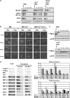
Figure 1
Figure 1
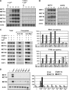
Cd2+inhibits the degradation-dependent regulation of Met4. (A) Cd2+ prevents MET gene repression by methionine in minimal medium. Total RNA was extracted at the indicated times after the addition of 1 mM L-methionine, in the presence (+Cd2+) or absence of 100 μM Cd2+, or in the presence of 100 μM of both Cd2+ and EGTA (+Cd2+, +EGTA), and MET gene expression was assessed by Northern analysis. (B) Cd2+ blocks elimination of endogenous GFP-Met4 by methionine. met4GFP-MET4 cells (strain CD240) were imaged at the indicated times after the addition (+Met) or not (−Met) of 1 mM L-methionine, in the presence (+Cd2+) or absence of 100 μM Cd2+, or in the presence of 100 μM of both Cd2+ and EGTA (+Cd2+, +EGTA). (C) Cd2+ stabilizes endogenous Met4 protein. met4HA3MET4 cells (strain CD233) were treated with (+Met) 1 mM L-methionine, in the presence (+Cd2+) or absence of 100 μM Cd2+, and total protein from TCA extracts was immunoblotted for the HA epitope or, as a control, lysyl-tRNA synthetase. (D) Met4p and TFIIB occupancy at MET promoters is elevated in response to Cd2+ exposure. A strain expressing both HA3Met4 and TFIIB9MYC (CD269) or an untagged isogenic control strain (W303-1A) were grown in B minimal medium and exposed to 1 mM L-methionine, in the presence or absence of 100 μM Cd2+. After crosslinking and immunoprecipitation with anti-HA or anti-Myc antibody, total DNA was analyzed by quantitative PCR with primer-pairs specific to the indicated Pol II promoters. ChIPs were performed at least twice and yielded similar results.
Figure 2
Figure 2
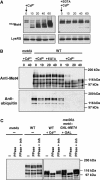
Cd2+ activates MET gene expression in rich medium. (A) Rapid induction of MET genes by Cd2+. MET gene expression in wild-type cells was assessed at the indicated times after the addition of 0.5 mM Cd2+ in the absence or presence of 0.5 mM EGTA. (B) Cd2+-mediated activation of MET genes depends on Met4. MET gene expression was assessed in wild-type and met4Δ (CC849-8A) cells at the indicated times after the addition of 0.5 mM Cd2+. (C) Met4 and TFIIB recruitment to MET gene promoters in response to Cd2+. ChIP experiments were carried out as in Figure 1 on wild-type cultures before and 40 min after the addition of 0.5 mM Cd2+. (D) Cd2+ specifically induces MET gene expression. MET gene expression was assessed in wild-type cells before and 40 min after addition of the indicated heavy metals by Northern analysis (left) and quantitative real-time RT–PCR (right). Heavy metals were added at the following concentrations: Cu2+ 1 mM, Zn2+ 1 mM, Co2+ 2 mM, Hg2+ 0.3 μM, Mn2+ 0.5 mM and Ag2+ 30 μM.
Figure 3
Figure 3
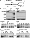
Cd2+ impairs SCFMet30-dependent ubiquitylation of Met4 in rich medium. (A) Collapse of Met4 isoforms in response to Cd2+. met4HA3MET4 cells (strain CD233) were exposed to 0.5 mM Cd2+ in the absence or presence of 0.5 mM EGTA and extracted proteins analyzed for Met4 and lysyl-tRNA synthetase abundance by immunoblot. (B) Loss of ubiquitinated Met4 species upon Cd2+ treatment. Endogenous Met4 was immunoprecipitated with anti-Met4 antibody and then immunoblotted with anti-Met4 and anti-ubiquitin antibodies. (C) Accumulation of phosphorylated Met4 species in response to Cd2+ and in the absence of Met30. Wild-type, met4Δ and met4GAL1-MET4, met30Δ strains were grown in rich medium containing 2% raffinose and harvested before and after 40 min of the indicated treatment with either 0.5 mM Cd2+ or galactose. Anti-Met4 immunoprecipitates were treated with lambda phosphatase in the presence or absence of phosphatase inhibitors and then immunoblotted with anti-Met4 antibody.
Figure 4
Figure 4
Cd2+ induces dissociation of the SCFMet30 ubiquitin ligase. (A) Met30 half-life is not altered by Cd2+ exposure. A strain bearing a GAL1-HA3MET30 construct was induced in galactose medium for 2 h, then transferred from galactose to 2% glucose- and 1 mM methionine-containing medium, in the presence or absence of 100 μM Cd2+, and HA3Met30 abundance assessed by anti-HA immunoblot at the indicated times after glucose repression. (B) The Met4–Met30 interaction is unaffected by Cd2+. An HA3Met30 fusion protein was expressed from a GAL1 promoter in B medium containing 2% galactose in the presence or absence of 100 μM Cd2+, Met4 immunoprecipitated from extracts with anti-Met4 antibody and immunoblotted with anti-Met4 and anti-HA antibodies. (C) The Skp1–Met30 interaction is specifically abolished by Cd2+ in minimal medium. HA3Met30 and HA3Cdc53 fusion proteins were expressed as in (B), extracts subjected to immunoprecipitation with the indicated antibodies and immunoblotted with anti-Skp1 and anti-HA antibodies. (D) The Skp1–Met30 interaction is specifically abolished upon Cd2+ exposure in rich medium. The experiments were performed as in (C) but cells were grown in rich medium and in the presence of 0.5 mM Cd2+. (E) The Skp1–Cdc4 interaction is not abolished upon Cd2+ exposure in rich medium. Met30MYC3 and Cdc4MYC3 fusion proteins were expressed from the constitutive CDC53 promoter (Ho et al, 2002) in rich medium in the presence or absence of 0.5 mM Cd2+ and protein interactions tested as in (C) with anti-MYC and anti-Skp1 antibodies. (F) Sic1 instability is not affected by Cd2+. A strain (CYS37) expressing a GFP-Sic1 fusion protein from the GAL1 promoter was grown in minimal medium, arrested in G1 phase by α-factor mating pheromone for 1.5 h, released into glucose medium in the presence and absence of 100 μM Cd2+, with or without α-factor, and GFP-Sic1 abundance assessed 120 min after the promoter shutoff by immunoblot with anti-GFP or, as a control, anti-lysyl-tRNA synthetase (LysRS) antibodies. (G) Quantification by flow cytometry of GFP-Sic stability in either an α-factor block or after release from the block in the presence and absence of 100 μM Cd2+. Cells were grown as described in (F) and the GFP-Sic1 fluorescent signal recorded and quantified at the indicated times using a Beckton Dickinson FacScalibur™ flow cytometer. (H) Met4 ubiquitylation by recombinant SCFMet30 is not affected by Cd2+. Purified recombinant Met4 and SCFMet30 produced in insect cells were incubated with E1 enzyme, Cdc34, ubiquitin and ATP, either in the absence or presence of the indicated amounts of Cd2+. Methyl-ubiquitin served as a control to demonstrate polyubiquitin chain formation on Met4.
Figure 5
Figure 5
Cd2+ triggers deubiquitylation of Met4 in rich medium in the absence of de novo Met4 synthesis. (A) Schematic representation of the experiment. (B) Cd2+-activated MET gene expression does not require de novo Met4 synthesis. A met4GAL1-MET4 strain (CC932-6D) was subjected to the regimen in (A) and assessed for MET gene expression at the indicated time points. (C) Cd2+ induces Met4 deubiquitylation. The same experiment as in (B) was performed except that Met4 was immunoprecipitated and immunoblotted with anti-Met4 antibody. (D) Inactivation of SCFMet30 does not induce Met4 deubiquitylation. Cultures of wild-type and cdc53-1 strains were shifted to the nonpermissive temperature of 37°C at the same time as repression of GAL1-MET4 by glucose. Cd2+ was either added or not at 60 min postshift.
Figure 6
Figure 6
Cd2+ sensitivity of met4Δ and met31Δ met32Δ strains. Serial dilutions of wild-type (WT, W303-1A), met28Δ (CC769-7D), met4Δ (CC849-1B), met31Δ (CC867-1C), met32Δ (CC845-1C) and met31Δ met32Δ (CC845-1A) strains were plated onto YNB minimal medium in the presence and absence of 20 μM Cd2+ and grown for 2 days.
Figure 7
Figure 7
Model for Cd2+-mediated activation of the MET gene network through the inhibition of the SCFMet30 ubiquitin ligase and activation of an uncharacterized deubiquitinylating enzyme (Ubp).

Similar articles

Cited by

References

    1. Aranda A, Del Olmo ML (2004) Exposure of Saccharomyces cerevisiae to acetaldehyde induces sulfur amino acid metabolism and polyamine transporter genes, which depend on Met4p and Haa1p transcription factors, respectively. Appl Environ Microbiol 70: 1913–1922 - PMC - PubMed
    1. Blackwell KJ, Tobin JM, Avery SV (1998) Manganese toxicity towards Saccharomyces cerevisiae: dependence on intracellular and extracellular magnesium concentrations. Appl Microbiol Biotechnol 49: 751–757 - PubMed
    1. Blaiseau PL, Isnard AD, Surdin-Kerjan Y, Thomas D (1997) Met31p and Met32p, two related zinc finger proteins, are involved in transcriptional regulation of yeast sulfur amino acid metabolism. Mol Cell Biol 17: 3640–3648 - PMC - PubMed
    1. Blaiseau PL, Thomas D (1998) Multiple transcription activation complexes tether the yeast activator Met4 to DNA. EMBO J 17: 6327–6336 - PMC - PubMed
    1. Cantoni GL (1977) S-adenosylmethionine: present status and future perspectives. In The Biochemistry of S-adenosylmethionine, Salvatore F, Borek E, Zappia V, Williams-Ashman HG, Schlenk F (eds) pp 557–577. New York: Columbia University Press

Publication types

MeSH terms