. 2005 Feb 9;24(3):543-53.
doi: 10.1038/sj.emboj.7600550. Epub 2005 Jan 20.

The DEAD box protein p68: a novel transcriptional coactivator of the p53 tumour suppressor

Affiliations

The DEAD box protein p68: a novel transcriptional coactivator of the p53 tumour suppressor

Gaynor J Bates et al. EMBO J. .

Abstract

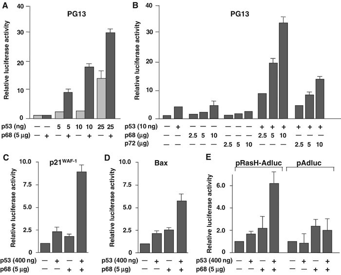
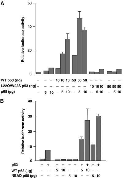
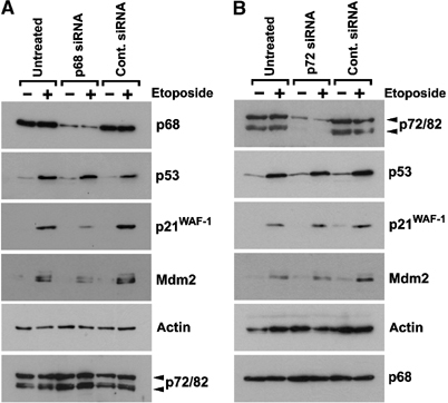
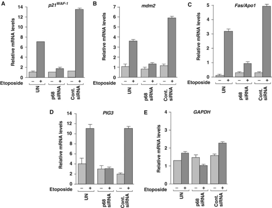
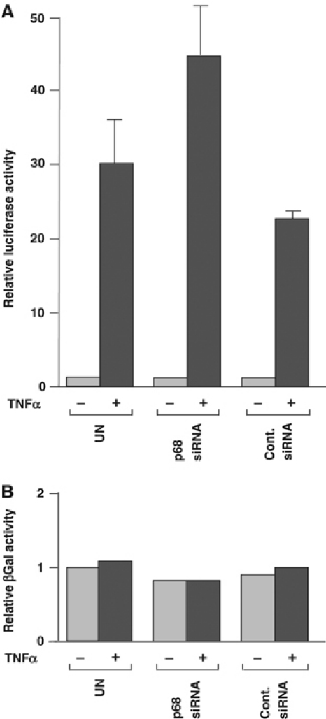
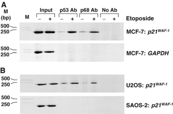
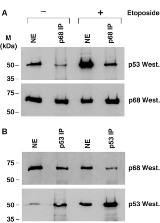
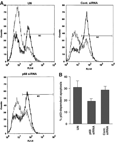
The DEAD box RNA helicase, p68, has been implicated in various cellular processes and has been shown to possess transcriptional coactivator function. Here, we show that p68 potently synergises with the p53 tumour suppressor protein to stimulate transcription from p53-dependent promoters and that endogenous p68 and p53 co-immunoprecipitate from nuclear extracts. Strikingly, RNAi suppression of p68 inhibits p53 target gene expression in response to DNA damage, as well as p53-dependent apoptosis, but does not influence p53 stabilisation or expression of non-p53-responsive genes. We also show, by chromatin immunoprecipitation, that p68 is recruited to the p21 promoter in a p53-dependent manner, consistent with a role in promoting transcriptional initiation. Interestingly, p68 knock-down does not significantly affect NF-kappaB activation, suggesting that the stimulation of p53 transcriptional activity is not due to a general transcription effect. This study represents the first report of the involvement of an RNA helicase in the p53 response, and highlights a novel mechanism by which p68 may act as a tumour cosuppressor in governing p53 transcriptional activity.


Figures

Figure 1
Figure 1
p68 stimulates p53 transcriptional activity from p53-responsive promoters. Effect of p68 on transactivation of the p53-responsive promoters PG13 (A, B), p21 (C), Bax (D) and pRasH-Adluc (E), fused to the luciferase reporter (pAdluc was used as a non-p53-responsive control (E)). In each case, the relative luciferase activity is shown with the basal activity of the promoter being taken as 1. Panels A and B show titres of the p53 and p68 plasmid DNAs, respectively, and the amounts used per ml of transfection mix are indicated. The amounts of reporter plasmid DNA used per ml of transfection mix were as follows: PG13, 2.5 μg; p21, 3 μg; Bax, 3 μg; pRasH-Adluc/pAdluc, 2.5 μg. Unless otherwise stated, the amounts of p53 plasmid transfected in these experiments had been optimised previously for the different promoters and were as follows: PG13, 10 ng; p21, Bax and pRasH-Adluc/pAdluc, 400 ng. Similarly, unless otherwise stated, 5 μg of p68 plasmid DNA was used. Graphs A and B represent the average results from two independent transfections, while graphs C–E represent average results from three independent experiments.
Figure 2
Figure 2
Coactivation requires transcriptionally active p53 but not helicase-active p68. (A) Coactivation of WT and L22Q/W23S (transcriptionally inactive) p53 transcriptional activity by p68. (B) Coactivation of WT p53 transcriptional activity by WT and NEAD (helicase-inactive) p68. Activity is determined by measurement of transactivation of the PG13 promoter fused to the luciferase reporter. The relative luciferase activity is shown, with the basal activity of the promoter being taken as 1. The amounts of p68 plasmid DNA used per ml of transfection mix are indicated and in all cases 10 ng of p53 plasmid DNA and 2.5 μg of PG13 reporter plasmid DNA were used per ml of transfection mix. Graphs represent the average results from two independent transfections.
Figure 3
Figure 3
p68 and p72 interact with p53 in vitro and in vivo. (A) Expression of GST vector control and GST-tagged p68/p72 used in the GST pull-down experiments as shown by Western blotting of cell lysates with a GST-specific antibody. (B) GST ‘pull-down' of in vitro-translated (35S-labelled) p53, showing both input and p53 species interacting with GST-tagged p68/p72. (C) Co-IP of p53 and p68 from nuclear extracts. p53 in U2OS extract was immunoprecipitated with the mouse monoclonal antibody (DO-1) and p68 and p53 in the IP were detected by Western blotting with rabbit polyclonal antibodies 2907 (p68) and CM1 (p53). (D) Reciprocal co-IP of p53 and p68. In this case, p68 was immunoprecipitated using the rabbit polyclonal antibody 2907 and immunoprecipitated p68 and p53 were detected by Western blotting with monoclonal antibodies PAb204 (p68) and DO-1 (p53). (E) Co-IP of p53 and p68 using a different p53-specific immunoprecipitating antibody. p53 in U2OS extract was immunoprecipitated with polyclonal antibody (CM1) and p68 and p53 were detected by Western blotting with monoclonal antibodies PAb204 (p68) and DO-1 (p53). (F) Co-IP of p53 and p72. Proteins immunoprecipitated by the p53 antibody (DO-1) (shown in (C)) were also Western blotted for p72 using the rabbit polyclonal antibody K14. (G) Reciprocal co-IP of p53 and p72. p72 was immunoprecipitated with the K14 antibody and p72 and p53 were detected by Western blotting with K14 and DO-1. Note that since only one p72 antibody is available, the same antibody had to be used for IP and Western blotting, giving a strong crossreaction with heavy chain (H). NE: nuclear extract; molecular weight markers (in kDa) are indicated. A nuclear extract from the p53-null cell line SAOS-2 and an irrelevant mouse or rabbit IgG (as appropriate; Cont. IP) were used as controls for IP.
Figure 4
Figure 4
RNAi depletion of p68, but not p72, inhibits expression of p53 target genes in response to DNA damage. Western blots showing expression of p68, p53, p21 and Mdm2 in MCF-7 cells, which had been transfected with (A) p68-specific or (B) p72-specific siRNA oligonucleotides. In both cases, a control siRNA was used and untransfected (UN) cells served as an additional control. In each case, the effect of treatment with the DNA-damaging agent etoposide (100 μM for 4 h) was examined. Equal amounts of protein (as determined by Bradford reagent (Sigma)) were loaded and detection of actin in the lysates was used as a loading control. Moreover, Western blots showing the levels of p68 and p72/82 in the reciprocal ‘knock-downs' confirm the specificity of the siRNAs. (The antibodies used for Western blotting are described in Materials and methods; 2907, K14 and DO-1 were used to detect p68, p72 and p53, respectively.)
Figure 5
Figure 5
RNAi depletion of p68 inhibits expression of cell cycle arrest and apoptosis-promoting p53 target genes. Quantitative RT–PCR of mRNA extracted from MCF-7 cells, which had been transfected with p68-specific siRNA. In both cases, a control siRNA was used and untransfected (UN) cells served as an additional control. Etoposide treatment was performed as in Figure 4. RT–PCR reactions to measure (A) p21, (B) mdm2, (C) Fas/APO1, (D) PIG3 and (E) GAPDH mRNA levels, in each case relative to β-actin, are shown. The average values from three independent RT–PCR reactions are shown.
Figure 6
Figure 6
RNAi depletion of p68 does not affect NF-κB activation by TNFα. (A) Induction of NF-κB activity by TNFα in cells transfected with p68 or control siRNA, compared with untransfected cells, as determined by measuring relative luciferase activity. The luciferase activity of untransfected cells, which had not been treated with TNFα, was taken as 1. (B) β-Galactosidase activity in cells. The levels shown represent values relative to that of untransfected and untreated cells, which were taken as 1. Graphs represent the average values from two independent experiments, and reactions were, in each case, performed in duplicate.
Figure 7
Figure 7
p68 is recruited to the p21 promoter, in a p53-dependent manner, in response to DNA damage. ChIP assays showing recruitment of p53 and p68 to the p21 promoter. (A) Enhancement of p53 and p68 recruitment to the p21 promoter in response to etoposide treatment. PCR reactions were performed using p21-specific and control (GAPDH-specific) primers to confirm specificity. (B) p68 to the p21 promoter in U2OS (p53 WT) but not in SAOS-2 (p53-null) cells. For all ChIP assays, samples of the input DNA, prior to IP with p53- or p68-specific antibodies (Ab), were used in the PCR reactions to confirm that equal amounts of DNA were present in the untreated and etoposide-treated samples. A control IP reaction with no antibody was also included. The cell line and the promoter-specific primers used for each assay are indicated as are DNA molecular weight markers.
Figure 8
Figure 8
DNA damage does not have a significant effect on the interaction between p68 and p53. Western blots showing co-IP of p68 and p53 from nuclear extracts of U2OS cells, which had been treated with 100 μM etoposide, with untreated cells as controls. (A) p68 IP. (B) p53 IP.
Figure 9
Figure 9
RNAi depletion of p68 causes a reduction in p53-dependent apoptosis. (A) Representative FACS profiles of SAOS-tetWTp53 transfected with a p68 siRNA or a control siRNA, with untransfected (UN) cells as a control. Cells were treated with 1 μM doxycycline for 36 h to induce p53 expression and thus apoptosis, with untreated cells acting as a control in each case. Apoptotic cells were stained as in Materials and methods and analysed by FACS. Graphs show number of cells (counts: y-axis) plotted against the fluorescence shift (FL1-H: x-axis). In each case, untreated cells are shown as a faint dotted line, while doxycycline-treated cells are shown as a bold solid line. (B) % apoptosis in doxycycline-treated cells minus % background apoptosis in untreated cells (i.e. p53-dependent apoptosis) for SAOS-tetWTp53 cells shown in (A). Graphs represent the average values from two independent experiments.

Similar articles

Cited by

References

    1. Balint EE, Vousden KH (2001) Activation and activities of the p53 tumour suppressor protein. Br J Cancer 85: 1813–1823 - PMC - PubMed
    1. Bond AT, Mangus DA, He F, Jacobson A (2001) Absence of Dbp2p alters both nonsense-mediated mRNA decay and rRNA processing. Mol Cell Biol 21: 7366–7379 - PMC - PubMed
    1. Causevic M, Hislop RG, Kernohan NM, Carey FA, Kay RA, Steele RJ, Fuller-Pace FV (2001) Overexpression and poly-ubiquitylation of the DEAD-box RNA helicase p68 in colorectal tumours. Oncogene 20: 7734–7743 - PubMed
    1. Courtois S, Verhaegh G, North S, Luciani MG, Lassus P, Hibner U, Oren M, Hainaut P (2002) DeltaN-p53, a natural isoform of p53 lacking the first transactivation domain, counteracts growth suppression by wild-type p53. Oncogene 21: 6722–6728 - PubMed
    1. Deguin-Chambon V, Vacher M, Jullien M, May E, Bourdon JC (2000) Direct transactivation of c-Ha-Ras gene by p53: evidence for its involvement in p53 transactivation activity and p53-mediated apoptosis. Oncogene 19: 5831–5841 - PubMed

Publication types

MeSH terms