. 2005 Jun 1;388(Pt 2):593-603.
doi: 10.1042/BJ20041517.

Interaction of U-box E3 ligase SNEV with PSMB4, the beta7 subunit of the 20 S proteasome

Affiliations

Interaction of U-box E3 ligase SNEV with PSMB4, the beta7 subunit of the 20 S proteasome

Marlies Löscher et al. Biochem J. .

Abstract

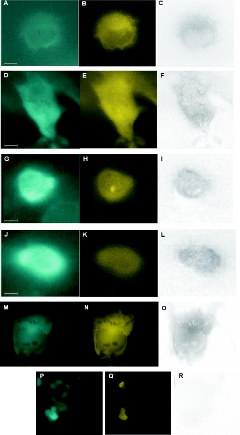
Recognition of specific substrates for degradation by the ubiquitin-proteasome pathway is ensured by a cascade of ubiquitin transferases E1, E2 and E3. The mechanism by which the target proteins are transported to the proteasome is not clear, but two yeast E3s and one mammalian E3 ligase seem to be involved in the delivery of targets to the proteasome, by escorting them and by binding to the 19 S regulatory particle of the proteasome. In the present study, we show that SNEV (senescence evasion factor), a protein with in vitro E3 ligase activity, which is also involved in DNA repair and splicing, associates with the proteasome by directly binding to the beta7 subunit of the 20 S proteasome. Upon inhibition of proteasome activity, SNEV does not accumulate within the cells although its co-localization with the proteasome increases significantly. Since immunofluorescence microscopy also shows increased co-localization of SNEV with ubiquitin after proteasome inhibition, without SNEV being ubiquitinated by itself, we suggest that SNEV shows E3 ligase activity not only in vitro but also in vivo and escorts its substrate to the proteasome. Since the yeast homologue of SNEV, Prp19, also interacts with the yeast beta7 subunit of the proteasome, this mechanism seems to be conserved during evolution. Therefore these results support the hypothesis that E3 ligases might generally be involved in substrate transport to the proteasome. Additionally, our results provide the first evidence for a physical link between components of the ubiquitin-proteasome system and the spliceosome.


Figures

Figure 1
Figure 1. SNEV and the β7 subunit of the proteasome are evolutionarily conserved
(A) Sequence comparison of the homologous proteins SNEV (H. sapiens), T10F2.4 (C. elegans) and Prp19 (S. cerevisiae). (B) Sequence comparison of the homologous β7 subunits of the 20 S proteasome PSMB4 (H. sapiens), pbs7 (C. elegans) and Pre4 (S. cerevisiae). Homology of SNEV and Prp19 by sequence comparison (BLAST database) revealed 23% (104/439) identities and 41% (184/439) positives; the last 81 (SNEV) or 99 (Prp19) amino acids were not matched. Homology of SNEV and T10F2.4 revealed 50% (253/505) identities and 67% (340/505) positives; the first 17 amino acids of T10F2.4 did not match. PSMB4 and Pre4 showed homology of 43% (100/232) identities and 64% (151/232) positives; the first 41 (PSMB4) or 29 (Pre4) amino acids are not included in the alignment. Homology of PSMB4 and pbs-7 revealed 33% (79/237) identities and 56% (134/237) positives; the first 41 (PSMB4) amino acids are not included in the alignment.
Figure 2
Figure 2. The interaction of SNEV and PSMB4 is evolutionarily highly conserved
(A) Directed Y2H assay for interaction of PRP19 and Pre4 as well as SNEV and PSMB4. The coding sequences were inserted in frame with the GAL4 AD and DNA BD respectively and co-transformed into S. cerevisiae AH109. Colony formation on 4× dropout medium was observed for SNEV(BD) and PSMB4(AD) (iv) in comparison with control co-transformations of SNEV(BD) and pGADT7 vector alone (v) and PSMB4(AD) and pGBKT7 vector alone (vi). The same result was seen for Prp19(AD) and Pre4(BD) (i) as opposed to the control experiments Pre4(BD) and pGADT7 vector, or Prp19(AD) and pGBKT7 vector (iii). (B, C) Y2H domain mapping of the interacting domains of SNEV and PSMB4. Y2H experiments with the deletion mutant constructs were performed as described in the text. Yeast colonies grown on 4× dropout medium (SD-4×) were re-streaked on SD-4×. ++, >80% of restreaked colonies grew again; −, <80% of restreaked colonies grew again; +, >80% of restreaked colonies grew again, but very slowly (smaller colonies were not visible until 14 days of incubation). (D) Additional Y2H experiments confirming the results of the domain mapping. (E) Calotte model of yeast Pre4 subunit structure (green) in the context of the proteasome (modified from protein database PDB accession no. 1FNT; Rasmol version 2.6). Putative interacting amino acids from the N-terminus of the mature subunit to amino acid 89 are coloured claret red. Putative interacting amino acids of the α-helix 1 region (amino acids 90–108), which are exposed to the surface are coloured: red, M93; blue, Q94; violet, E97; cyan, R98; orange, K101; dark green, D102; purple, V104; black, T105; yellow, A108. Since the α-helix 1 is highly conserved, we presume that these amino acids are also exposed in the mammalian proteasome, (F) surface model of the bovine β7 subunit incorporated into the proteasome (modified from protein database PDB accession no. 1IRU). Stereo pictures with Swiss PDB viewer version 3.7, peptides coloured as in Figure 2(C).
Figure 3
Figure 3. Interaction of in vitro translated SNEV and Prp19 with the β7 subunit of the proteasome
(A) SNEV and PSMB4 were in vitro translated as fusions to the Myc- and HA-tag respectively and labelled with 35S-methionine. Precipitation of Myc-tagged SNEV by anti-Myc antibody-coupled beads resulted in co-precipitation of HA-tagged PSMB4 as revealed by SDS/PAGE and autoradiography. Lane 1, negative control, HA–PSMB4 on c-Myc beads; lane 2, CoIP of Myc–SNEV and HA–PSMB4; lane 3, input of in vitro translated HA–PSMB4; lane 4, input of in vitro translated Myc–SNEV in co-precipitation. (B) Pre4, in vitro translated as fusion to the c-Myc-tag, and Prp19–His6, recombinantly expressed in yeast, were incubated together, followed by incubation with Ni2+-NTA beads. Beads were washed and SDS/PAGE was performed. Precipitated Myc–Pre4 was detected with anti-c-Myc antibody. Lane 1, CoIP of Prp19–His6 and Myc–Pre4, lane 2, Myc–Pre4 on Ni2+-NTA beads as negative control and lane 3, input of in vitro translated Myc–Pre4 in CoIP.
Figure 4
Figure 4. The interaction of SNEV and PSMB4 is direct
Purified GST–PSMB4 fusion protein expressed in E. coli was incubated together with affinity-purified His6–SNEV, expressed by baculovirus/insect cell system and precipitated on GST–Sepharose beads. The precipitate was separated by SDS/PAGE and detected by Western blot. SNEV was detected by anti-His4 antibody. As control, GST and GST–PSMB4 were detected with anti-GST antibody. Lane 1, CoIP of GST–PSMB4 (without propeptide, 50 kDa) and His6–SNEV (56 kDa) on GST-beads; lane 2, CoIP of GST (26 kDa) alone and His6–SNEV on GST-beads [image acquired by LumiImager (Roche); since the GST signal is too strong, it is displayed as inverted]; lane 3, His6–SNEV alone on GST-beads.
Figure 5
Figure 5. SNEV interacts with the proteasome in vivo but is not degraded by it
(A) Proteasome was precipitated from HEK-293 cell lysate by anti-α2-coupled Protein A beads. After washing the beads and elution of proteins by resuspending the beads in SDS-loading buffer, SDS/PAGE was performed. Precipitation of the proteasome was confirmed on Western blots with anti-PSMB4 antibody, whereas co-precipitated SNEV was detected with anti-SNEV antibody. Lane 1, mouse-anti-α2 antibody-coupled beads co-precipitate PSMB4 and SNEV; lane 2, mouse-anti-Myc antibody-coupled beads were used as negative control, cell lysate on anti-c-Myc-coupled beads; lane 3, whole cell lysate as input control. (B) Western blot of HeLa cell extracts that were treated with 25 μM MG132 for 6 h (+) versus untreated controls (−). Protein amounts of SNEV and β-actin do not change, indicating that SNEV is not a substrate to the proteasome. To control the effect of MG132 on the cells, we probed for p53, a known proteasomal substrate, and for polyubiquinated proteins, using anti-p53 and anti-ubiquitin antibodies respectively. Indeed, both p53 and ubiquitinated proteins accumulate on proteasome inhibition. (C) In vitro 26 S proteasome degradation assay. SNEV was incubated with or without the 26 S proteasome for 0 and 2 h in an in vitro degradation assay. Quantification of SNEV protein amounts detected on Western blots was achieved by densitometry. As positive control for the in vitro degradation assay, IκBα was used as the model substrate and detected in a Western blot by anti-IκBα antibody. Although only 80% of SNEV was detected after incubation for 2 h at 37 °C, this degradation was not dependent on the addition of the 26 S proteasome, suggesting that SNEV is not a substrate of the proteasome.
Figure 6
Figure 6. Interaction of SNEV and PSMB4 can be visualized in cells by FRET
COS-1 (AF) and HEK-293 cells (GL) were co-transfected with plasmids carrying SNEV, N-terminally fused to ECFP (cyan), and PSMB4, C-terminally fused to EYFP (yellow). A, D, G, J: microscopic picture using CFP-filter (SNEV); B, E, H, K: picture using YFP-filter (PSMB4) and C, F, I, L: calculated net FRET signal of SNEV and PSMB4. Scale bar, 10 μm (×60 objective). MO: Positive control using an EYFP–ECFP fusion protein in COS-1 cells. PR: Negative control using HEK-293 cells co-transfected with plasmids coding for ECFP–Δ66-SNEV and PSMB4–EYFP fusion proteins. The pictures (PR) were taken using a lower magnification (×40 objective).
Figure 7
Figure 7. Partial nuclear co-localization of SNEV with the proteasome and with ubiquitin in HeLa cells after MG132 treatment
HeLa cells were treated with DMSO as control (AC) and with 25 μM MG132 (dissolved in DMSO) for 6 h. Indirect immunofluorescence was performed using anti-SNEV (green, A, D) and anti-α2 proteasome subunit antibodies (red, B, E). Overlay of these pictures shows partial co-localization (yellow) of SNEV and the proteasome, which is rare in untreated (C, yellow spot indicated by arrow) versus proteasome-inhibited cells (F). Co-localization was also observed between SNEV (G, green) and ubiquitin (H, red), as can be seen in the overlay (I), although only in MG 132-treated cells. (J) Representative DAPI staining for visualization of the nucleus. Scale bar, 5 μm.

References

    1. Ciechanover A., Schwartz A. L. The ubiquitin-proteasome pathway: the complexity and myriad functions of proteins death. Proc. Natl. Acad. Sci. U.S.A. 1998;95:2727–2730. - PMC - PubMed
    1. Unno M., Mizushima T., Morimoto Y., Tomisugi Y., Tanaka K., Yasuoka N., Tsukihara T. The structure of the mammalian 20 S proteasome at 2.75 Å resolution. Structure. 2002;10:609–618. - PubMed
    1. Ferrell K., Wilkinson C. R., Dubiel W., Gordon C. Regulatory subunit interactions of the 26 S proteasome, a complex problem. Trends Biochem. Sci. 2000;25:83–88. - PubMed
    1. Weissman A. M. Themes and variations on ubiquitylation. Nat. Rev. Mol. Cell. Biol. 2001;2:169–178. - PubMed
    1. Grillari J., Hohenwarter O., Grabherr R. M., Katinger H. Subtractive hybridization of mRNA from early passage and senescent endothelial cells. Exp. Gerontol. 2000;35:187–197. - PubMed

Publication types

MeSH terms