

In vitro assembled, recombinant infectious bronchitis viruses demonstrate that the 5a open reading frame is not essential for replication
- PMID: 15661153
- PMCID: PMC7111797
- DOI: 10.1016/j.virol.2004.10.045
In vitro assembled, recombinant infectious bronchitis viruses demonstrate that the 5a open reading frame is not essential for replication
Abstract
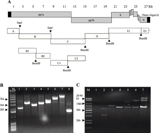
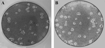
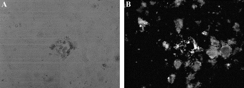
Molecular clones of infectious bronchitis virus (IBV), derived from the Vero cell adapted Beaudette strain, were constructed, using an in vitro assembly method. In vitro transcribed RNA from a cDNA template that had been constructed from seven cDNA fragments, encompassing the entire genome of IBV, was electroporated into BHK-21 cells. The cells were overlaid onto the susceptible Vero cells and viable virus was recovered from the molecular clone. The molecularly cloned IBV (MIBV) demonstrated growth kinetics, and plaque size and morphology that resembled the parental Beaudette strain IBV. The recombinant virus was further manipulated to express enhanced green fluorescent protein (EGFP) by replacing an open reading frame (ORF) of the group-specific gene, ORF 5a, with the EGFP ORF. The rescued recombinant virus, expressing EGFP (GIBV), replicated to lower viral titers and formed smaller plaques compared to the parental virus and the MIBV. After six passages of GIBV, a minority of plaques were observed that had reverted to the larger plaque size and virus from these plaques no longer expressed EGFP. Direct sequencing of RT-PCR products derived from cells infected with the plaque-purified virus, which had lost expression of EGFP, confirmed loss of the EGFP ORF. The loss of EGFP expression (Delta5a IBV) was also accompanied by reversion to growth kinetics resembling the standard virus and intact recombinant virus. This study demonstrates that the 5a ORF is not essential for viral multiplication in Vero cells.
Figures
Similar articles
-
Replication and packaging of coronavirus infectious bronchitis virus defective RNAs lacking a long open reading frame.J Virol. 1996 Dec;70(12):8660-8. doi: 10.1128/JVI.70.12.8660-8668.1996. J Virol. 1996. PMID: 8970992 Free PMC article.
-
Utilizing fowlpox virus recombinants to generate defective RNAs of the coronavirus infectious bronchitis virus.J Gen Virol. 2000 Dec;81(Pt 12):2855-2865. doi: 10.1099/0022-1317-81-12-2855. J Gen Virol. 2000. PMID: 11086116
-
[Expression of green fluorescent protein using an infectious cDNA clone of infectious bronchitis virus].Bing Du Xue Bao. 2011 Jan;27(1):11-7. Bing Du Xue Bao. 2011. PMID: 21462501 Chinese.
-
The S2 Subunit of Infectious Bronchitis Virus Beaudette Is a Determinant of Cellular Tropism.J Virol. 2018 Sep 12;92(19):e01044-18. doi: 10.1128/JVI.01044-18. Print 2018 Oct 1. J Virol. 2018. PMID: 30021894 Free PMC article.
-
Multiple novel non-canonically transcribed sub-genomic mRNAs produced by avian coronavirus infectious bronchitis virus.J Gen Virol. 2020 Oct;101(10):1103-1118. doi: 10.1099/jgv.0.001474. J Gen Virol. 2020. PMID: 32720890 Free PMC article.
Cited by
-
Avian infectious bronchitis virus (AIBV) review by continent.Front Cell Infect Microbiol. 2024 Feb 5;14:1325346. doi: 10.3389/fcimb.2024.1325346. eCollection 2024. Front Cell Infect Microbiol. 2024. PMID: 38375362 Free PMC article. Review.
-
Differential localization and turnover of infectious bronchitis virus 3b protein in mammalian versus avian cells.Virology. 2006 Feb 20;345(2):337-45. doi: 10.1016/j.virol.2005.09.069. Epub 2005 Nov 18. Virology. 2006. PMID: 16298409 Free PMC article.
-
Amino acid residues critical for RNA-binding in the N-terminal domain of the nucleocapsid protein are essential determinants for the infectivity of coronavirus in cultured cells.Nucleic Acids Res. 2006;34(17):4816-25. doi: 10.1093/nar/gkl650. Epub 2006 Sep 13. Nucleic Acids Res. 2006. PMID: 16971454 Free PMC article.
-
Generating antibodies to the gene 3 proteins of infectious bronchitis virus.Methods Mol Biol. 2008;454:163-89. doi: 10.1007/978-1-59745-181-9_14. Methods Mol Biol. 2008. PMID: 19057877 Free PMC article.
-
Acquisition of cell-cell fusion activity by amino acid substitutions in spike protein determines the infectivity of a coronavirus in cultured cells.PLoS One. 2009 Jul 2;4(7):e6130. doi: 10.1371/journal.pone.0006130. PLoS One. 2009. PMID: 19572016 Free PMC article.
References
Publication types
MeSH terms
Substances
Grants and funding
LinkOut - more resources
Full Text Sources
Other Literature Sources
