

Canine distemper virus-induced depletion of uninfected lymphocytes is associated with apoptosis
- PMID: 15661329
- PMCID: PMC7127010
- DOI: 10.1016/j.vetimm.2004.09.032
Canine distemper virus-induced depletion of uninfected lymphocytes is associated with apoptosis
Abstract
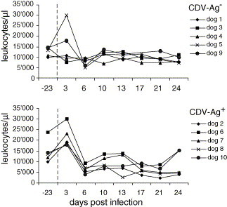
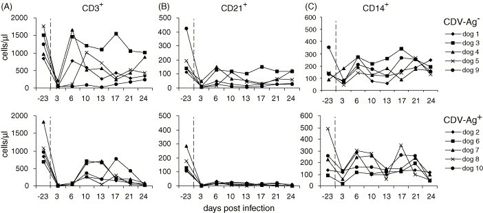
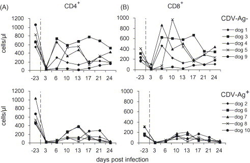
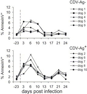
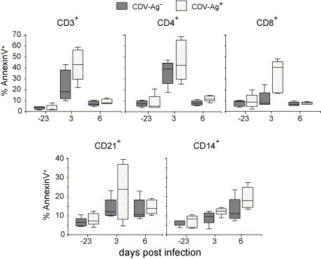
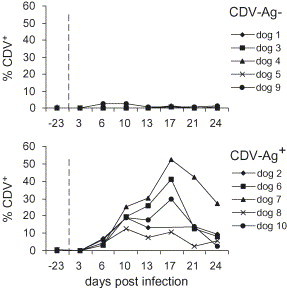
Canine distemper virus (CDV), a negative stranded RNA morbillivirus, causes a multisystemic disease in dogs, which is associated with a severe immune suppression. The aim of the study was to examine the influence of early CDV infection on leukocyte depletion, lymphopenia and virus-induced cell death in dogs infected with a virulent CDV strain. From 10 infected dogs, peripheral blood leukocytes were harvested periodically, phenotyped and analyzed for CDV antigen content and apoptosis using Annexin V-FITC and propidium iodide labeling. CDV infection induced a severe CD3+ T cell and CD21+ B cell depletion in all animals at 3 days post-infection (d.p.i.). For dogs with severe distemper, developing virus persistence in the lymphoid tissue and central nervous system, this lymphopenia lasted until the end of the experiment. Increased levels of lymphocyte apoptosis were found at 3 d.p.i., and monocyte apoptosis at 6 d.p.i. This was more prominent in the group of animals with severe distemper. At 3 d.p.i. no leukocyte infection was detectable indicating that the early lymphocyte depletion and apoptosis was not a direct consequence of virus infection. Taken together, our results demonstrate that CDV-induced lymphopenia is an early event and that the degree of lymphocyte depletion correlates with the severity of disease and virus persistence in the lymphoid tissue and central nervous system.
Figures
Similar articles
-
Partial protection and intrathecal invasion of CD8(+) T cells in acute canine distemper virus infection.Vet Microbiol. 2001 Nov 26;83(3):189-203. doi: 10.1016/s0378-1135(01)00422-9. Vet Microbiol. 2001. PMID: 11574169
-
Phenotypical characterization of T and B cell areas in lymphoid tissues of dogs with spontaneous distemper.Vet Immunol Immunopathol. 2000 Jan 31;73(1):83-98. doi: 10.1016/s0165-2427(99)00156-7. Vet Immunol Immunopathol. 2000. PMID: 10678401
-
Comparison of canine distemper virus strains in gnotobiotic dogs: effects on lymphoid tissues.Am J Vet Res. 1977 Dec;38(12):1919-22. Am J Vet Res. 1977. PMID: 304324
-
Pathogenesis and immunopathology of systemic and nervous canine distemper.Vet Immunol Immunopathol. 2009 Jan 15;127(1-2):1-18. doi: 10.1016/j.vetimm.2008.09.023. Epub 2008 Oct 4. Vet Immunol Immunopathol. 2009. PMID: 19019458 Review.
-
Establishment of central nervous system infection by canine distemper virus: breach of the blood-brain barrier and facilitation by antiviral antibody.Vet Immunol Immunopathol. 1987 Dec;17(1-4):471-82. doi: 10.1016/0165-2427(87)90163-2. Vet Immunol Immunopathol. 1987. PMID: 2963430 Review.
Cited by
-
Membrane fusion-mediated autophagy induction enhances morbillivirus cell-to-cell spread.J Virol. 2012 Aug;86(16):8527-35. doi: 10.1128/JVI.00807-12. Epub 2012 May 30. J Virol. 2012. PMID: 22647692 Free PMC article.
-
Ferrets as a model for morbillivirus pathogenesis, complications, and vaccines.Curr Top Microbiol Immunol. 2009;330:73-87. doi: 10.1007/978-3-540-70617-5_4. Curr Top Microbiol Immunol. 2009. PMID: 19203105 Free PMC article. Review.
-
Immunodeficiencies caused by infectious diseases.Vet Clin North Am Small Anim Pract. 2010 May;40(3):409-23. doi: 10.1016/j.cvsm.2010.01.006. Vet Clin North Am Small Anim Pract. 2010. PMID: 20471525 Free PMC article. Review.
-
Comparative Immunopathology of Cetacean morbillivirus Infection in Free-Ranging Dolphins From Western Mediterranean, Northeast-Central, and Southwestern Atlantic.Front Immunol. 2019 Mar 18;10:485. doi: 10.3389/fimmu.2019.00485. eCollection 2019. Front Immunol. 2019. PMID: 30936878 Free PMC article.
-
Hematological Changes and Immunomodulation of Neutrophil and Monocyte Populations in Shelter Dogs.Animals (Basel). 2025 Jul 6;15(13):1988. doi: 10.3390/ani15131988. Animals (Basel). 2025. PMID: 40646887 Free PMC article.
References
-
- Appel M.J., Pearce-Kelling S., Summers B.A. Dog lymphocyte cultures facilitate the isolation and growth of virulent canine distemper virus. J. Vet. Diagn. Invest. 1992;4:258–263. - PubMed
-
- Appel M.J. Pathogenesis of canine distemper. Am. J. Vet. Res. 1969;30:1167–1182. - PubMed
-
- Bautista E.M., Ferman G.S., Golde W.T. Induction of lymphopenia and inhibition of T cell function during acute infection of swine with foot and mouth disease virus (FMDV) Vet. Immunol. Immunopathol. 2003;92:61–73. - PubMed
-
- Bollo E., Zurbriggen A., Vandevelde M., Fankhauser R. Canine distemper virus clearance in chronic inflammatory demyelination. Acta Neuropathol. (Berl.) 1986;72:69–73. - PubMed
MeSH terms
Substances
LinkOut - more resources
Full Text Sources
