Review
. 2005 Feb;17(1):35-46.
doi: 10.1016/j.ceb.2004.12.009.

Kinetochore-spindle microtubule interactions during mitosis

Affiliations
Review

Kinetochore-spindle microtubule interactions during mitosis

Susan L Kline-Smith et al. Curr Opin Cell Biol. 2005 Feb.

Abstract

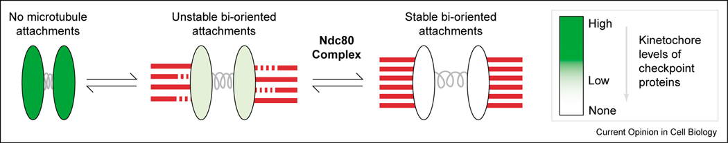
The kinetochore is a proteinaceous structure that assembles onto centromeric DNA and mediates chromosome attachment to microtubules during mitosis. This description is deceivingly simple: recent proteomic studies suggest that the diminutive kinetochores of Saccharomyces cerevisiae are comprised of at least 60 proteins organized into as many as 14 different subcomplexes. Many of these proteins, such as the centromeric histone variant CENP-A, and entire subcomplexes, such as the Ndc80(Hec1) complex, are conserved from yeast to humans despite the diverse nature of the DNA sequences on which they assemble. There have recently been advances in our understanding of the molecular basis of how kinetochores establish dynamic attachments to spindle microtubules, and how these attachments are correctly oriented to ensure segregation of sister chromatids to daughter cells.


Figures

Figure 1
Figure 1
Formation of stable kinetochore–microtubule attachments. Microtubules are in red and centromeric chromatin is in gray. Kinetochores are indicated by ovals in various shades of green corresponding to the level of kinetochore-localized spindle checkpoint proteins, which are progressively depleted as microtubules attach to kinetochores. With 95% depletion of Ndc80 complex subunits, significant depletion of checkpoint proteins still occurs, suggesting the existence of some type of attachment between kinetochores and spindle microtubules. However, stable kinetochore fibers do not form, and chromosome alignment and segregation is severely perturbed. Thus, the Ndc80 complex plays a critical role in stabilization of microtubule attachments, allowing the formation of mature kinetochore fibers capable of aligning and segregating chromosomes properly.
Figure 2
Figure 2
Regulation of kinetochore–microtubule dynamics. The assembly dynamics of kinetochore-attached microtubules is coupled to chromosome movement on the spindle. Regulators of microtubule dynamics localized to kinetochores, such as the CLASP family of MAPs and the kinesin-13 family of microtubule depolymerases, are likely to play important roles in this process. Kinetochore-localized CLASPs (in green) may stimulate the growth of kinetochore-attached microtubules during anti-poleward movements that align chromosomes at the metaphase plate. Polymerization of kinetochore microtubule plus ends is also necessary for poleward microtubule flux during metaphase. In contrast, the microtubule depolymerase activity of kinesin-13 proteins (in blue) may contribute to poleward chromosome movement, although discrepancies between studies in different systems concerning this putative role need to be resolved. Arrows indicate the predicted direction of chromosome movement. MT, microtubule.
Figure 3
Figure 3
Error correction mechanisms ensure chromosome bi-orientation on the spindle. Attachment of sister kinetochores to microtubules emanating from opposing spindle poles, a configuration referred to as bi-orientation, is critical to ensure chromosome alignment and segregation. This cartoon depicts how errors in microtubule attachments may be resolved in vertebrate cells by Aurora B kinase and the kinesin-13 protein MCAK, a newly identified substrate of Aurora B. Kinetochores and centromeric heterochromatin are in gray, unphosphorylated MCAK (active microtubule depolymerase) is in green, and MCAK phosphorylated in the neck region (inactive depolymerase) is in black. Throughout mitosis, populations of both phosphorylated and unphosphorylated MCAK exist at the centromere, although the precise localization of active versus inactive MCAK is controversial. One interpretation is that active MCAK is more prevalent when incorrect attachments are present and tension is low. MCAK can then depolymerize inappropriately attached microtubules. Upon attachment of sister kinetochores to opposite spindle poles, the resulting increase in tension prevents Aurora-B-mediated re-orientation, perhaps by affecting local regulation of MCAK activity. In fungi, the Dam1 complex is the key target of Aurora BIpl1 implicated in this error correction process. Such a complex has not been identified in metazoans.

Similar articles

Cited by

References

    1. McIntosh JR, Grishchuk EL, West RR: Chromosome-microtubule interactions during mitosis. Annu Rev Cell Dev Biol 2002, 18:193–219. - PubMed
    1. Cleveland DW, Mao Y, Sullivan KF: Centromeres and kinetochores: from epigenetics to mitotic checkpoint signaling. Cell 2003, 112:407–421. - PubMed
    1. Lew DJ, Burke DJ: The spindle assembly and spindle position checkpoints. Annu Rev Genet 2003, 37:251–282. - PubMed
    1. Amor DJ, Kalitsis P, Sumer H, ChooKH: Building the centromere: from foundation proteins to 3D organization. Trends Cell Biol 2004, 14:359–368. - PubMed
    1. McAinsh AD, Tytell JD, Sorger PK: Structure, function, and regulation of budding yeast kinetochores. Annu Rev Cell Dev Biol 2003, 19:519–539. - PubMed

Publication types

LinkOut - more resources