

Experimental autoimmune encephalomyelitis in the rat spinal cord: lesion detection with high-resolution MR microscopy at 17.6 T
- PMID: 15661692
- PMCID: PMC7975015
Experimental autoimmune encephalomyelitis in the rat spinal cord: lesion detection with high-resolution MR microscopy at 17.6 T
Abstract
Background and purpose: Experimental autoimmune encephalomyelitis (EAE) is an inflammatory demyelinating disorder of the CNS and an animal model of multiple sclerosis. We used high-field MR microscopy at 17.6 T to image spinal cord inflammatory lesions in the acute stage of chronic relapsing rat EAE. We sought to compare lesions detected on MR imaging with histopathologic findings and to quantify the inflammatory lesion load.
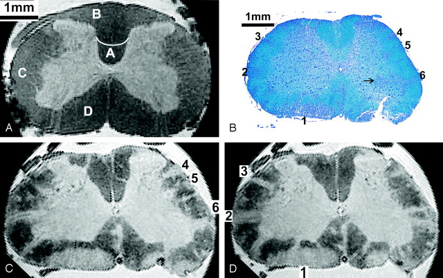
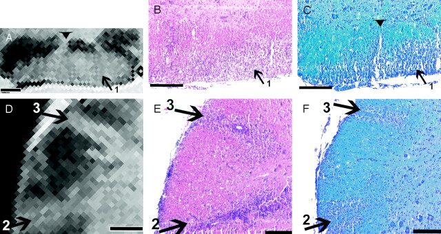
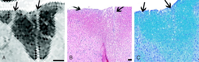
Methods: Imaging of fixed spinal cord specimens was performed by using a 3D gradient-echo sequence with a spatial resolution of 35 x 35 x 58 microm3 and a total imaging time of 5.5 hours. Histopathologic analysis was performed by staining axial sections with hematoxylin-eosin or Luxol fast blue to identify cellular infiltration and demyelination.
Results: Clinical signs of EAE occurred on days 10-14 after immunization. On day 22, healthy white matter and gray matter were differentiated by high contrast on T2*-weighted images, with white matter lesions appearing as hyperintense areas in the normal-appearing white matter. Inflammatory lesions identified on histopathologic evaluation were readily detected with MR imaging and vice versa. MR imaging and histopathologic analysis had excellent correlation regarding the extent of white matter lesions. Inflammatory infiltrates of gray matter were not detectable with MR imaging. Using a semiautomatic segmentation of the acquired MR data, we could quantify white matter lesion load.
Conclusion: Ex vivo high-resolution MR microscopy of the spinal cord at 17.6 T allows rapid and highly accurate determination of CNS inflammation by demonstrating virtually all histologically detectable white matter inflammatory lesions.
Figures


References
-
- McDonald WI, Compston A, Edan G, et al. Recommended diagnostic criteria for multiple sclerosis: guidelines from the International Panel on the Diagnosis of Multiple Sclerosis. Ann Neurol 2001;50:121–127 - PubMed
-
- Filippi M, Dousset V, McFarland HF, et al. Role of magnetic resonance imaging in the diagnosis and monitoring of multiple sclerosis: consensus report of the White Matter Study group. J Magn Reson Imaging 2002;15:499–504 - PubMed
-
- Lycklama a Nijeholt G, Barkhof F. Differences between subgroups of MS: MRI findings and correlation with histopathology. J Neurol Sci 2003;206:173–174 - PubMed
-
- Filippi M, Grossman RI. MRI techniques to monitor MS evolution: the present and the future. Neurology 2002;58:1147–1153 - PubMed
-
- Lucchinetti C, Bruck W, Parisi J, et al. Heterogeneity of multiple sclerosis lesions: implications for the pathogenesis of demyelination. Ann Neurol 2000;47:707–717 - PubMed
Publication types
MeSH terms
LinkOut - more resources
Full Text Sources
Medical