

Intradural spinal vein enlargement in craniospinal hypotension
- PMID: 15661695
- PMCID: PMC7975033
Intradural spinal vein enlargement in craniospinal hypotension
Abstract
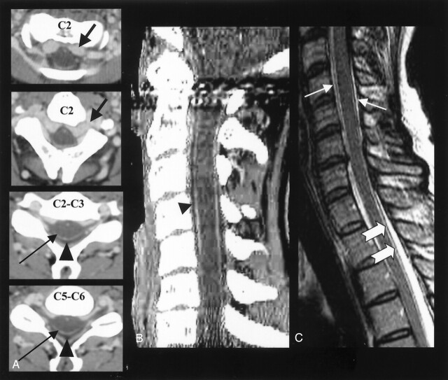
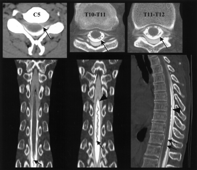
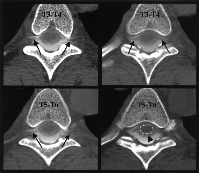
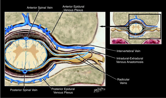
We present a case of craniospinal hypotension in a 45-year-old woman with an associated epidural pseudomeningocele extending the entire length of the spine. The epidural pseudomeningocele was caused by a CSF leak at the T8 level. In addition to typical low-pressure symptoms, the epidural pseudomeningocele caused atypical symptoms characterized by positional thoracic radiculopathy. Craniospinal hypotension was associated with massive cervical epidural venous engorgement, as well as enlargement of the posterior spinal cord vein, which was reminiscent of a dural arteriovenous fistula at CT myelography. Enlargement of the posterior spinal vein is explained by the Monro-Kellie hypothesis, and the spinal analog to enlarged cerebral veins known to be associated with intracranial hypotension.
Figures
Comment in
-
"Epidural" vertebral venous plexus.AJNR Am J Neuroradiol. 2006 Jan;27(1):7; author reply 7. AJNR Am J Neuroradiol. 2006. PMID: 16418345 Free PMC article. No abstract available.
Similar articles
-
Intracranial hypotension as a cause of radiculopathy from cervical epidural venous engorgement: case report.AJNR Am J Neuroradiol. 2002 Apr;23(4):618-21. AJNR Am J Neuroradiol. 2002. PMID: 11950654 Free PMC article.
-
Angiographic features of spontaneous intracranial hypotension.AJNR Am J Neuroradiol. 2003 Apr;24(4):704-6. AJNR Am J Neuroradiol. 2003. PMID: 12695207 Free PMC article.
-
[Epidural varicosis as a rare cause of acute radiculopathy with complete foot paresis--case report and literature review].Z Orthop Ihre Grenzgeb. 2007 Jan-Feb;145(1):55-60. doi: 10.1055/s-2007-960503. Z Orthop Ihre Grenzgeb. 2007. PMID: 17345544 Review. German.
-
CSF hypovolemia vs intracranial hypotension in "spontaneous intracranial hypotension syndrome".Neurology. 2003 Mar 25;60(6):941-7. doi: 10.1212/01.wnl.0000049933.51044.81. Neurology. 2003. PMID: 12654957
-
[Intracranial dural fistula with spinal cord venous drainage. Apropos of 2 cases].J Neuroradiol. 1994 Apr;21(2):134-54. J Neuroradiol. 1994. PMID: 8014658 Review. French.
Cited by
-
Cerebrospinal fluid leakage and headache after lumbar puncture: a prospective non-invasive imaging study.Brain. 2015 Jun;138(Pt 6):1492-8. doi: 10.1093/brain/awv016. Epub 2015 Feb 13. Brain. 2015. PMID: 25688077 Free PMC article.
-
Neuroimaging in superficial siderosis: an in-depth look.AJNR Am J Neuroradiol. 2010 Jan;31(1):5-14. doi: 10.3174/ajnr.A1628. Epub 2009 Sep 3. AJNR Am J Neuroradiol. 2010. PMID: 19729538 Free PMC article. Review.
-
Epidural blood patch at C2: diagnosis and treatment of spontaneous intracranial hypotension.AJNR Am J Neuroradiol. 2005 Nov-Dec;26(10):2663-6. AJNR Am J Neuroradiol. 2005. PMID: 16286420 Free PMC article.
-
Spinal perimedullary vein enlargement sign: an added value for the differentiation between intradural-extramedullary and intramedullary tumors on magnetic resonance imaging.Neuroradiology. 2016 Nov;58(11):1117-1124. doi: 10.1007/s00234-016-1744-4. Epub 2016 Sep 5. Neuroradiology. 2016. PMID: 27596484
-
Intradural venous engorgement of CSF-venous fistula mimics spinal dural arteriovenous fistula on MRI: A novel case report and review of literature.Neuroradiol J. 2025 Jun 13:19714009251351292. doi: 10.1177/19714009251351292. Online ahead of print. Neuroradiol J. 2025. PMID: 40514049 Free PMC article.
References
-
- Schaltenbrand G. Neuere Anschauungen zur Pathophysiologic der Liquorzikulation. Zentralbl Neurochir 1938;3:290–300
-
- Schaltenbrand G. Normal and pathological physiology of cerebrospinal fluid circulation. Lancet 1953;1:805–809 - PubMed
-
- Schievink WI. Spontaneous spinal cerebrospinal fluid leaks: a review. Neurosurg Focus 2000;9:1–9 - PubMed
-
- Chung SJ, Kim JS, Lee MC. Syndrome of cerebral spinal fluid hypovolemia: clinical and imaging features and outcome. Neurology 1993;43:609–611 - PubMed
-
- Christofordis GA, Mehta BA, Landi JL, et al. Spontaneous intracranial hypotension: report of four cases and review of the literature. Neuroradiology 1998;40:636–643 - PubMed
Publication types
MeSH terms
LinkOut - more resources
Full Text Sources
Medical