

MR quantitation of volume and diffusion changes in the developing brain
- PMID: 15661698
- PMCID: PMC7975043
MR quantitation of volume and diffusion changes in the developing brain
Abstract
Background and purpose: Brain volume and diffusion change during maturation. Quantitation of these changes may be helpful in understanding normal brain development. We used diffusion-weighted imaging to characterize the volumetric and diffusion changes in vivo.
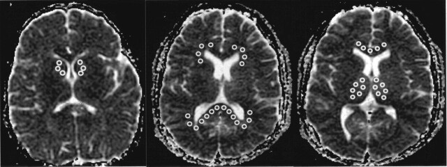
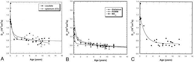
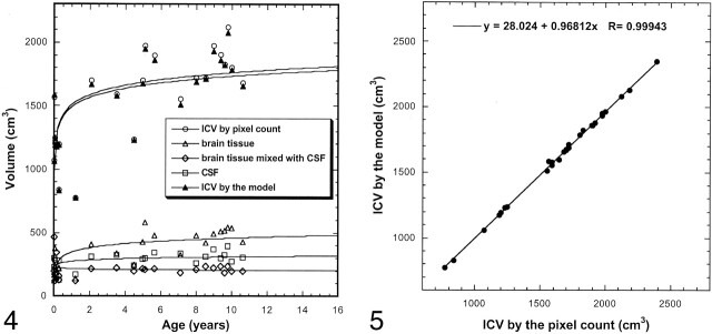
Methods: We recruited 30 pediatric volunteers (aged 1 month-17 years; 14 male, 16 female). Diffusion was measured in three orthogonal directions with a b value of 1000 s/mm2. The diffusion parameters from the entire brain were calculated and fitted to a triple gaussian model. In addition, region-of-interest measurements were made in caudate, thalamus, genu and splenium of the corpus callosum, and periventricular white matter (PVWM). The brain volume was measured by counting pixels and by using the model.
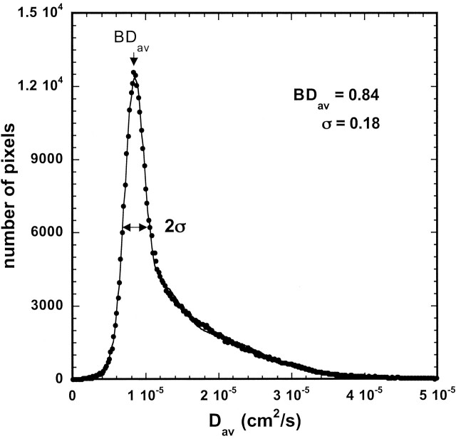
Results: Water diffusion of the whole brain, caudate, thalamus, genu and splenium of the corpus callosum, and PVWM decreased during maturation, with the most significant change within the first 2 years. Robust negative correlations were found between age and the measured average diffusion constant (Dav) values in each of the measured locations (P <.005). Volumes of different cerebral compartments and the total intracranial volume (ICV) increased rapidly during the first 2 years of life and then had a slower growth process through adolescence. Age was correlated with the ICV and the volume of each brain compartment (P <.005).
Conclusion: Brain diffusion decreases and brain volume increases during maturation, with the most significant changes occurring within the first 2 years of life. The brain model used in this study provides a good estimate of the increasing brain volume.
Figures
Similar articles
-
Diffusion-tensor MR imaging of gray and white matter development during normal human brain maturation.AJNR Am J Neuroradiol. 2002 Oct;23(9):1445-56. AJNR Am J Neuroradiol. 2002. PMID: 12372731 Free PMC article.
-
Diffusion changes in patients with systemic lupus erythematosus.Magn Reson Imaging. 2007 Apr;25(3):399-405. doi: 10.1016/j.mri.2006.09.037. Epub 2006 Nov 29. Magn Reson Imaging. 2007. PMID: 17371731
-
Diffusion tensor imaging assessment of brain white matter maturation during the first postnatal year.AJR Am J Roentgenol. 2007 Aug;189(2):476-86. doi: 10.2214/AJR.07.2132. AJR Am J Roentgenol. 2007. PMID: 17646476
-
Sexual dimorphism of brain developmental trajectories during childhood and adolescence.Neuroimage. 2007 Jul 15;36(4):1065-73. doi: 10.1016/j.neuroimage.2007.03.053. Epub 2007 Apr 6. Neuroimage. 2007. PMID: 17513132 Free PMC article.
-
Normal brain maturation during childhood: developmental trends characterized with diffusion-tensor MR imaging.Radiology. 2001 Nov;221(2):349-58. doi: 10.1148/radiol.2212001702. Radiology. 2001. PMID: 11687675
Cited by
-
Volumetric apparent diffusion coefficient histogram analysis in term neonatal asphyxia treated with hypothermia.Br J Radiol. 2024 Jun 18;97(1159):1302-1310. doi: 10.1093/bjr/tqae105. Br J Radiol. 2024. PMID: 38775658 Free PMC article.
-
Optic radiation structure and anatomy in the normally developing brain determined using diffusion MRI and tractography.Brain Struct Funct. 2015 Jan;220(1):291-306. doi: 10.1007/s00429-013-0655-y. Epub 2013 Oct 30. Brain Struct Funct. 2015. PMID: 24170375 Free PMC article.
-
Developmental score of the infant brain: characterizing diffusion MRI in term- and preterm-born infants.Brain Struct Funct. 2020 Nov;225(8):2431-2445. doi: 10.1007/s00429-020-02132-4. Epub 2020 Aug 17. Brain Struct Funct. 2020. PMID: 32804327 Free PMC article.
-
Progressive frontal morphology changes during the first year of a modified Pi procedure for scaphocephaly.Childs Nerv Syst. 2016 Feb;32(2):337-44. doi: 10.1007/s00381-015-2914-0. Epub 2015 Sep 26. Childs Nerv Syst. 2016. PMID: 26409882
-
Advanced imaging in paediatric neuroradiology.Pediatr Radiol. 2009 Jun;39 Suppl 3:456-63. doi: 10.1007/s00247-009-1230-9. Pediatr Radiol. 2009. PMID: 19440766 Review. No abstract available.
References
-
- Smart JL, Dobbing J. Vulnerability of developing brain, IX: the effect of nutritional growth retardation on the timing of the brain growth-spurt. Biol Neonate 1971;19:363–378 - PubMed
-
- Uluğ AM. Monitoring brain development with quantitative diffusion imaging. Dev Sci 2002;5:286–292
-
- Huppi PS, Maier SE, Eeled S, et al. Microstructural development of human newborn cerebral white matter assessed in vivo by diffusion tensor magnetic resonance imaging. Pediatr Res 1998;44:584–590 - PubMed
Publication types
MeSH terms
Grants and funding
LinkOut - more resources
Full Text Sources
Medical