

Case Reports
Malformations of cortical development: high-resolution MR and diffusion tensor imaging of fiber tracts at 3T
Affiliations
- PMID: 15661702
- PMCID: PMC7975027
Item in Clipboard
Case Reports
Malformations of cortical development: high-resolution MR and diffusion tensor imaging of fiber tracts at 3T
AJNR Am J Neuroradiol.
2005 Jan.
Abstract
Patients with malformations of cortical development and epilepsy may have a variety of abnormal brain findings, including abnormal gyral patterns, cortical thickening, decreased volume of white matter, and increased diffusion of white matter. The status of individual white matter fiber tracts, however, is unknown. We present a case of bilateral frontal schizencephaly and subcortical heterotopia and illustrate alterations of white matter fascicles by combined structural and functional diffusion tensor imaging at 3 T.
Figures
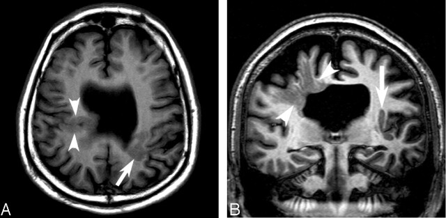
Conventional MR images obtained at 1.5 T and 3 T. A, Axial T1-weighted image obtained at 1.5 T shows bilateral closed lip schizencephaly (arrow and arrowheads) and absent septum pellucidum. B, Coronal image obtained at 3T through the schizencephalic cleft reveals individual linear radiations of gray matter signal (arrowheads) extending from the cortex to the ventricle.
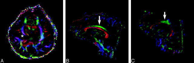
Principal eigenvector maps obtained from diffusion tensor imaging. A, Axial color map corresponding to the main direction of the white matter fiber orientation: by convention, blue indicates fibers (e.g., corona radiata) running in the superior to inferior direction, green indicates anterior to posterior (e.g., superior longitudinal fasciculus), and red indicates right-left fibers (e.g., corpus callosum). In our patient, the white matter fibers show deviation in the main direction of the diffusion tensor around the abnormal gray matter of the schizencephalic cleft (asterisk). B, In a healthy individual, the cingulum (arrow) is a long thin fascicle, lying immediately superior to the corpus callosum, oriented in an anterioposterior direction. C, The cingulum in our patient (arrow) is shortened and distorted. Note also thinning of the corpus callosum (red) below the cingulum.
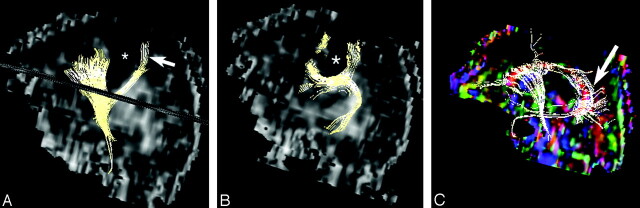
Fiber tract reconstruction images. A, 2D projections of fiber tract reconstruction superimposed on sagittal fractional anisotropy map showing the deep schizencephaly (asterisk) separating the corticospinal tract (arrow) from other fibers. B, In our patient, the superior longitudinal fasciculus is poorly developed and deviated by the schizencephalic cleft (asterisk). C, Only a few thin fibers are seen in the in the body of the corpus callosum, and the fibers in the splenium (arrow) are oriented in complex directions.
Similar articles
-
Diffusion tensor imaging in presymptomatic and early Huntington's disease: Selective white matter pathology and its relationship to clinical measures.Mov Disord. 2006 Sep;21(9):1317-25. doi: 10.1002/mds.20979. Mov Disord. 2006. PMID: 16755582
-
Subcortical pathways serving cortical language sites: initial experience with diffusion tensor imaging fiber tracking combined with intraoperative language mapping.Neuroimage. 2004 Feb;21(2):616-22. doi: 10.1016/j.neuroimage.2003.09.047. Neuroimage. 2004. PMID: 14980564 Free PMC article.
-
Exploring white matter tracts in band heterotopia using diffusion tractography.Ann Neurol. 2002 Sep;52(3):327-34. doi: 10.1002/ana.10295. Ann Neurol. 2002. PMID: 12205645
-
Abnormalities of gyration, heterotopias, tuberous sclerosis, focal cortical dysplasia, microdysgenesis, dysembryoplastic neuroepithelial tumour and dysgenesis of the archicortex in epilepsy. Clinical, EEG and neuroimaging features in 100 adult patients.Brain. 1995 Jun;118 ( Pt 3):629-60. doi: 10.1093/brain/118.3.629. Brain. 1995. PMID: 7600083 Review.
-
White matter anatomy: what the radiologist needs to know.Neuroimaging Clin N Am. 2013 May;23(2):197-216. doi: 10.1016/j.nic.2012.12.002. Epub 2013 Jan 20. Neuroimaging Clin N Am. 2013. PMID: 23608685 Review.
Cited by
-
Characterizing White Matter Tract Organization in Polymicrogyria and Lissencephaly: A Multifiber Diffusion MRI Modeling and Tractography Study.AJNR Am J Neuroradiol. 2020 Aug;41(8):1495-1502. doi: 10.3174/ajnr.A6646. Epub 2020 Jul 30. AJNR Am J Neuroradiol. 2020. PMID: 32732266 Free PMC article.
-
Loss of electrical anisotropy is an unrecognized feature of dystrophic muscle that may serve as a convenient index of disease status.Clin Neurophysiol. 2016 Dec;127(12):3546-3551. doi: 10.1016/j.clinph.2016.09.017. Epub 2016 Oct 13. Clin Neurophysiol. 2016. PMID: 27825055 Free PMC article.
-
Diffusion tensor imaging of midline posterior fossa malformations.Pediatr Radiol. 2006 Jun;36(6):510-7. doi: 10.1007/s00247-006-0146-x. Epub 2006 Apr 21. Pediatr Radiol. 2006. PMID: 16708205
-
Imaging surgical epilepsy in children.Childs Nerv Syst. 2006 Aug;22(8):786-809. doi: 10.1007/s00381-006-0132-5. Epub 2006 Jul 13. Childs Nerv Syst. 2006. PMID: 16838193 Review.
-
Functional and resting-state characterizations of a periventricular heterotopic nodule associated with epileptogenic activity.Neurosurg Focus. 2020 Feb 1;48(2):E10. doi: 10.3171/2019.11.FOCUS19765. Neurosurg Focus. 2020. PMID: 32006947 Free PMC article.
References
-
- Yakovlev PI, Wadsworth RC. Schizencephalies: a study of the congenital clefts in the cerebral mantle. 1. Clefts with fused lips. J Neuropathol Exp Neurol 1946;5:116–130 - PubMed
-
- Barkovich AJ, Norman D. MR imaging of schizencephaly. AJR Am J Roentgenol 1988;150:1391–1396 - PubMed
-
- Eriksson SH, Rugg-Gunn FJ, Symms MR, et al. Diffusion tensor imaging in patients with epilepsy and malformations of cortical development. Brain 2001;124:617–626 - PubMed
-
- Sisodiya SM. Wiring, dysmorphogenesis and epilepsy: a hypothesis. Seizure 1995;4:169–185 - PubMed
Publication types
MeSH terms
LinkOut - more resources
Full Text Sources
Medical