

Parapharyngeal neuroglial heterotopia presenting as a growing single locular cyst: MR imaging findings
- PMID: 15661709
- PMCID: PMC7975011
Parapharyngeal neuroglial heterotopia presenting as a growing single locular cyst: MR imaging findings
Abstract
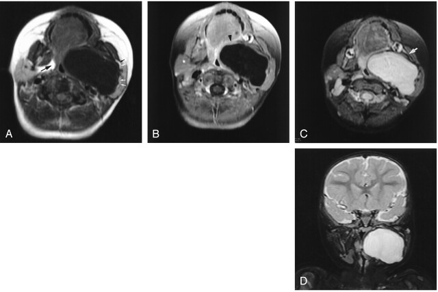
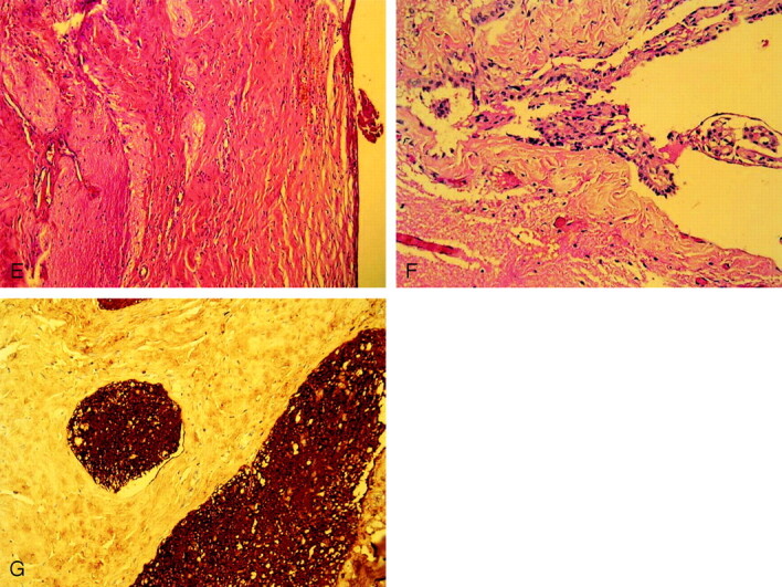
Heterotopic brain presenting as a giant, growing, single locular cyst at the parapharyngeal space has not been reported before, to our knowledge. We present such a case, with MR imaging findings, in a 13-month-old girl. A well-demarcated giant cystic mass was noted in the left parapharyngeal space from the skull base to the submandibular region. Airway compression and deformity of the left mandible with subluxation of the temporomandibular joint were noted. The cyst contained a clear fluid that was isointense to CSF with all pulse sequences. Wall enhancement was noted on contrast-enhanced T1-weighted images. No connection to intracranial structures was noted. Histologic findings were compatible with neuroglial heterotopia.
Figures
Similar articles
-
Extensive parapharyngeal and skull base neuroglial ectopia; a challenge for differential diagnosis and treatment: case report.Sao Paulo Med J. 2010;128(5):302-5. doi: 10.1590/s1516-31802010000500011. Sao Paulo Med J. 2010. PMID: 21181072 Free PMC article.
-
Parapharyngeal Neuroglial Heterotopia: A Case Report and Literature Review.Am J Case Rep. 2020 Nov 6;21:e926300. doi: 10.12659/AJCR.926300. Am J Case Rep. 2020. PMID: 33156818 Free PMC article. Review.
-
Parapharyngeal neuroglial heterotopia extending through the skull base in a neonate with airway obstruction.J Pediatr Surg. 2007 Oct;42(10):1764-7. doi: 10.1016/j.jpedsurg.2007.06.014. J Pediatr Surg. 2007. PMID: 17923212
-
Case series of congenital heterotopic neuroglial tissue in the parapharyngeal space.Int J Pediatr Otorhinolaryngol. 2016 Jul;86:77-81. doi: 10.1016/j.ijporl.2016.04.026. Epub 2016 Apr 26. Int J Pediatr Otorhinolaryngol. 2016. PMID: 27260585
-
Parapharyngeal neuroglial heterotopia appearing as high uptake on 18F-FDG PET: case report and literature review of radiographical findings.Acta Neurochir (Wien). 2018 Apr;160(4):801-809. doi: 10.1007/s00701-017-3403-x. Epub 2017 Dec 2. Acta Neurochir (Wien). 2018. PMID: 29197937 Review.
Cited by
-
Rare tumors in pediatric age group: Single center experience from Saudi Arabia.Rare Tumors. 2021 Feb 24;13:2036361321997331. doi: 10.1177/2036361321997331. eCollection 2021. Rare Tumors. 2021. PMID: 33708364 Free PMC article.
-
An unusual presentation of neuroglial heterotopia: case report.BJR Case Rep. 2020 Sep 29;6(2):20190116. doi: 10.1259/bjrcr.20190116. eCollection 2020 Sep. BJR Case Rep. 2020. PMID: 33029378 Free PMC article.
-
Extensive parapharyngeal and skull base neuroglial ectopia; a challenge for differential diagnosis and treatment: case report.Sao Paulo Med J. 2010;128(5):302-5. doi: 10.1590/s1516-31802010000500011. Sao Paulo Med J. 2010. PMID: 21181072 Free PMC article.
-
Respiratory difficulty caused by an ectopic brain tissue mass in the neck of a two-month-old baby: a case report.J Med Case Rep. 2011 Jun 8;5:220. doi: 10.1186/1752-1947-5-220. J Med Case Rep. 2011. PMID: 21649939 Free PMC article.
-
Parapharyngeal Neuroglial Heterotopia: A Case Report and Literature Review.Am J Case Rep. 2020 Nov 6;21:e926300. doi: 10.12659/AJCR.926300. Am J Case Rep. 2020. PMID: 33156818 Free PMC article. Review.
References
-
- Azumi N, Matsuno T, Tateyama M, Inoue K. So-called nasal glioma. Acta Pathol Jpn 1984;34:215–220 - PubMed
-
- Hendrikson M, Faye-Petersen O, Johnson DG. Cystic and solid heterotopic brain in the face and neck: a review and report of an unusual case. J Pediatr Surg 1990;25:766–768 - PubMed
-
- Broniatowski M, Witt WJ, Shah AC, et al. Glial tissue in the parapharyngeal space. Arch Otolaryngol 1981;107:638–641 - PubMed
-
- Forte V, Friedberg J, Thorner P, Park A. Heterotopic brain in the parapharyngeal space. Int J Pediatr Otorhinolaryngol 1996;37:253–260 - PubMed
-
- Behar PM, Muller S, Gerber ME, Todd NM. Heterotopic neurotopic neuroglial tissue causing airway obstruction in the newborn. Arch Otolaryngol Head Neck Surg 2001;127:997–1002 - PubMed
Publication types
MeSH terms
LinkOut - more resources
Full Text Sources
Medical