

Case Reports
MR imaging findings of uveal leiomyoma: three cases
Affiliations
- PMID: 15661710
- PMCID: PMC7975025
Item in Clipboard
Case Reports
MR imaging findings of uveal leiomyoma: three cases
AJNR Am J Neuroradiol.
2005 Jan.
Abstract
Leiomyoma is a rare tumor arising from the uveal tract. Fundoscopically, it appears as a yellowish-white, elevated mass and cannot be readily distinguished from melanoma or other uveal tumors. Cross-sectional imaging may have an important role, particularly when the opaque ocular media or a vitreous hemorrhage precludes a clear ophthalmoscopic view. In this respect, radiologists should be aware of suggestive findings of uveal leiomyoma to avoid an incorrect diagnosis and unnecessary radical surgery. We report MR imaging findings of three cases of uveal leiomyoma.
Figures
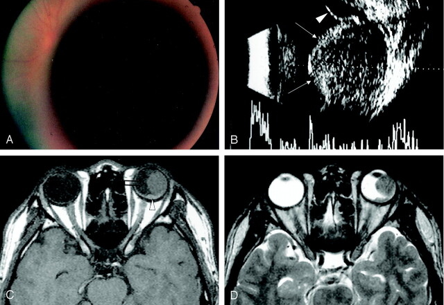
A 24-year-old man with sudden decrease of left visual acuity. A, Fundus photograph shows a dark, large, circumscribed, view-obstructing mass. B, Transocular sonography reveals a well-marginated endophytic mass with low internal reflectivity (arrows). Subretinal effusion is also observed (arrowhead). C, The mass located at the ciliochoroidal region is large, protuberant, and isointense to brain on a precontrast T1-weighted axial image (open arrow). Small amount of subretinal hemorrhage is noted (arrowhead). D, T2-weighted fast spin-echo axial image also shows an intraocular mass isointense to brain.
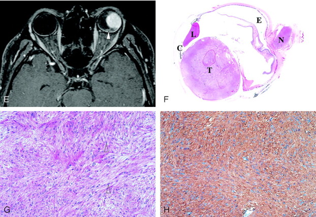
(continued) E, On gadolinium-enhanced T1-weighted axial image, a homogeneous, enhancing mass is seen at the ciliochoroidal region (open arrow), as is a small amount of nonenhancing subretinal hemorrhage around the posterior pole (arrowhead). F, Low-magnification photograph of a cut specimen shows a well-circumscribed ciliochoroidal mass involving the whole stroma of the choroids (hematoxylin and eosin staining; magnification ×2). C indicates ciliary body; E, subretinal effusion; L, lens; N, optic nerve; and T, tumor. G and H, Hematoxylin and eosin staining (G) shows spindle cells arranged in intersecting fascicles, and tumor cells that have cigar-shaped nuclei with blunted end (arrowheads). The chromatin pattern is vesicular and mitotic figures are not seen. The tumor cells are positively stained for smooth muscle-specific actin (H).
Similar articles
-
Observations on intraocular leiomyomas.Trans Pa Acad Ophthalmol Otolaryngol. 1990;42:945-50. Trans Pa Acad Ophthalmol Otolaryngol. 1990. PMID: 2084991 Review.
-
Posterior choroidal leiomyoma: a rare case report and literature review.APMIS. 2015 Jun;123(6):540-5. doi: 10.1111/apm.12384. Epub 2015 Apr 23. APMIS. 2015. PMID: 25907891 Review.
-
Choroidal hemangioma: MR findings and differentiation from uveal melanoma.AJNR Am J Neuroradiol. 1998 Sep;19(8):1441-7. AJNR Am J Neuroradiol. 1998. PMID: 9763374 Free PMC article.
-
MR imaging findings of the uveal schwannoma.AJNR Am J Neuroradiol. 2009 Apr;30(4):769-73. doi: 10.3174/ajnr.A1467. Epub 2009 Jan 22. AJNR Am J Neuroradiol. 2009. PMID: 19164439 Free PMC article.
-
Leiomyoma in the posterior choroid: a case report.J Korean Med Sci. 2002 Jun;17(3):429-33. doi: 10.3346/jkms.2002.17.3.429. J Korean Med Sci. 2002. PMID: 12068155 Free PMC article.
Cited by
-
T2 Fluid-Attenuated Inversion Recovery Imaging of Uveal Melanomas and Other Ocular Pathology.Ocul Oncol Pathol. 2016 Oct;2(4):251-261. doi: 10.1159/000447265. Epub 2016 Jul 1. Ocul Oncol Pathol. 2016. PMID: 27843906 Free PMC article.
-
Magnetic Resonance Imaging in the Clinical Care for Uveal Melanoma Patients-A Systematic Review from an Ophthalmic Perspective.Cancers (Basel). 2023 May 30;15(11):2995. doi: 10.3390/cancers15112995. Cancers (Basel). 2023. PMID: 37296958 Free PMC article. Review.
-
Growth of congenital malignant teratoid medulloepithelioma of the ciliary body: a case study.J Neurooncol. 2010 Feb;96(3):443-8. doi: 10.1007/s11060-009-9982-5. Epub 2009 Aug 8. J Neurooncol. 2010. PMID: 19669095
-
MR features of intraocular ectopic lacrimal tissue.AJNR Am J Neuroradiol. 2006 Nov-Dec;27(10):2196-8. AJNR Am J Neuroradiol. 2006. PMID: 17110692 Free PMC article.
References
-
- McMahon RT. Anatomy, congenital anomalies, and tumors. In: Peyman CA, Sanders DR, Goldberg MF, eds. Principles and practice of ophthalmology. Philadelphia: Sauders;1980. :1491–1553
-
- Lemke AJ, Hosten N, Bornfeld N, et al. Uveal melanoma: correlation of histopathologic and radiologic findings by using thin-section MR imaging with a surface coil. Radiology 1999;210:775–783 - PubMed
-
- Shields JA, Shields CL. Observations on intraocular leiomyomas. Trans Pa Acad Ophthalmol Otolaryngol 1990;42:945–950 - PubMed
-
- Shields JA, Shields CL, Eagle RC Jr, et al. Observations on seven cases of intraocular leiomyoma. Arch Ophthalmol 1994;112:521–528 - PubMed
-
- Osterlind A. Trends in incidence of ocular malignant melanoma in Denmark 1943–1982. Int J Cancer 1987;40:161–164 - PubMed
Publication types
MeSH terms
Substances
LinkOut - more resources
Full Text Sources
Medical