. 2005 Jan 20;433(7023):269-77.
doi: 10.1038/nature03206.

Structure of human follicle-stimulating hormone in complex with its receptor

Affiliations

Structure of human follicle-stimulating hormone in complex with its receptor

Qing R Fan et al. Nature. .

Abstract

Follicle-stimulating hormone (FSH) is central to reproduction in mammals. It acts through a G-protein-coupled receptor on the surface of target cells to stimulate testicular and ovarian functions. We present here the 2.9-A-resolution structure of a partially deglycosylated complex of human FSH bound to the extracellular hormone-binding domain of its receptor (FSHR(HB)). The hormone is bound in a hand-clasp fashion to an elongated, curved receptor. The buried interface of the complex is large (2,600 A2) and has a high charge density. Our analysis suggests that all glycoprotein hormones bind to their receptors in this mode and that binding specificity is mediated by key interaction sites involving both the common alpha- and hormone-specific beta-subunits. On binding, FSH undergoes a concerted conformational change that affects protruding loops implicated in receptor activation. The FSH-FSHR(HB) complexes form dimers in the crystal and at high concentrations in solution. Such dimers may participate in transmembrane signal transduction.


Conflict of interest statement

Competing interests statement The authors declare that they have no competing financial interests.

Figures

Figure 1
Figure 1
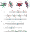
Crystal structure of human FSH bound to FSHFHB a, b, Ribbon diagram of the complex structure shown in two views related by a 90° rotation about the vertical axis. FSH α-chains and β-chains are in green and cyan, respectively. FSHRHB is in red. The observed N-linked carbohydrates at N52 and N78 of FSH-α, N7 and N24 of FSH-β, and N191 of FSHRHB are in yellow. Disulphide bonds are in black. c, Schematic diagram of the topology of FSHRHB structure. β-Strands are shown as arrows. Red represents strands located at the concave face of FSHRHB; pink represents strands on the convex face.
Figure 2
Figure 2
Recognition of FSH by FSHRHB. a, The left panel shows the molecular surface of FSHRHB with the imprint of bound FSH (Cα trace in green for α- and cyan for β-chains). Coloured patches represent FSHRHB residues that are buried at the receptor—hormone interface by FSH-α alone (green), by FSH-β alone (cyan), or both FSH-α and FSH-β (magenta). The right panel shows the molecular surface of FSH with the imprint of FSHRHB (Cα trace in red). Green denotes residues of FSH-α buried by FSHRHB; cyan represents buried FSH-β residues. b, Electrostatic potential surface of FSHRHB (left) and FSH (right). Residues of FSHRHB are marked red; residues of the FSH-α and FSH-β chains are in green and cyan, respectively. c–e, Sequence alignment. Secondary structure assignments are shown as arrows for β-strands and cylinders for α-helices. For each residue buried at the receptor—ligand interface or the receptor dimer interface, the fractional solvent accessibility is indicated by an open circle if it is greater than 0.4, a half-filled circle if it is 0.1–0.4, and a filled circle if it is less than 0.1. Residues implicated in binding specificity are marked by asterisks. c, Human FSHR, LHR and TSHR sequences in the region of the hormone-binding domain. β-Strands located at the concave face of FSHR are coloured red, whereas strands on the convex face are in pink. FSHRHB residues buried at the receptor—ligand interface by FSH-α alone (green), FSH-β alone (cyan), or both FSH-α and FSH-β (magenta) are highlighted. FSHRHB residues buried at the receptor dimer interface are boxed in orange. N-linked glycosylation is marked by a black triangle. d, Human FSH, CG, LH and TSH β-chain sequences. FSH-β residues buried at the receptor—ligand interface are highlighted in cyan. e, The common human α-chain sequence. FS-Hα residues buried at the receptor—ligand interface are highlighted in green.
Figure 3
Figure 3
Interactions at the receptor—ligand interface. a, Ribbon diagram showing the top view of the FSH—FSHRHB complex. The view in the right panel is tilted to highlight the regions of FSHRHB (red), and FSH-α (green) and FSH-β (cyan) chains that are involved in direct contacts at the receptor—ligand interface. Dashed circles mark the locations of L55 and K179 in the FSHRHB structure. b, Detailed views of the interactions at the specificity pockets for L55 and K179 of FSHRHB. The dotted molecular surface of FSH is shown in cyan in each panel. c, Close-up stereo view of the interactions between the C-terminal region of FSH-α and FSHRHB.
Figure 4
Figure 4
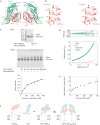
Different conformations in free and receptor-bound FSH. a, Cα trace superposition of four independent copies of FSH. One protomer of FSH is bound to the FSHRHB shown in red; its α-chain is in green and β-chain in cyan. The second copy of receptor-bound FSH is in blue. The two protomers of free FSH are in orange and pink. b, Stereo diagram of the superposition of one receptor-bound FSH (α-chain in green; β-chain cyan) with one free hormone (orange). The view is rotated 90° about the vertical axis from a. Every 20th residue of the receptor-bound FSH is marked. Red highlights the segment of the βℒ2 loop that is involved in receptor binding.
Figure 5
Figure 5
Dimer of the FSH—FSHRHB complex. a, Ribbon diagram of a non-crystallographic dimer of the FSH—FSHRHB complex (FSH—FSHRHB and FSH’—FSHRHB’). The receptor is in red, and the α- and β-chains of the hormone are in green and cyan, respectively. b, Stereo view of the direct contacts (distances <4Å) observed at the dimer interface. c, Crosslinking of the FSH—FSHRHB complex by glutaraldehyde. The SDS gel contains the FSH—FSHRHB control (lane 2) and 120 μM FSH—FSHRHB complex reacted with 120 μM glutaraldehyde (lane 3). The native gel contains the FSH—FSHRHB control (lane 1), and FSH—FSHRHB complex reacted with glutaraldehyde at a 1:1 molar ratio and at concentrations of 30, 60,120,180, 240, 300 and 360 μM (lanes 2–8, respectively). Densities from the dimer bands on native gels as a function of protein concentration were fitted to the dimerization equation with a single Kd (453 ± 140 μM) for six experiments, with two repeated trials for each molar ratio of FSH—FSHRHB to glutaraldehyde (3:1, 3:2, 1:1). The average dimer fraction at each protein concentration is plotted together with the fitted curve. d, Analytical ultracentrifugation analysis of dimer formation by the FSH— FSHRHB complex. Shown are equilibrium sedimentation data (green open circle) taken at 12,000 r.p.m. with a loading concentration of 1.05 mg ml−1, and the best-fit curve (black solid line through the data) for a monomer—dimer reversible equilibrium model. Under the fit, the theoretical sum is deconvoluted into contributions from the monomer (blue line) and dimer (red line) species, respectively. Residuals in absorbance units at 280 nm are shown at the top of the plot. e, Apparent hydrodynamic radii of FSH—FSHRHB measured by dynamic light scattering at concentrations of 15, 27, 50, 75, 100, 150, 200, 270, 300, 360 and 390 μM. f, Model for FSHR activation by FSH. Projections of the FSH—FSHRHB complex and bovine rhodopsin structures are used to represent the hormone (cyan), the hormone-binding domain (orange) and the linker and transmembrane domain (black dotted trace) of the receptor. The αℒ1 and aL3 loops of FSH are proposed to contact the transmembrane domain directly.

Comment in

Similar articles

Cited by

References

    1. Dias JA, et al. Molecular, structural, and cellular biology of follitropin and follitropin receptor. Vitam Horm. 2002;64:249–322. - PubMed
    1. Simoni M, Gromoll J, Nieschlag E. The follicle-stimulating hormone receptor: biochemistry, molecular biology, physiology, and pathophysiology. Endocr Rev. 1997;18:739–773. - PubMed
    1. Themmen APN, Huhtaniemi IT. Mutations of gonadotropins and gonadotropin receptors: elucidating the physiology and pathophysiology of pituitary-gonadal function. Endocr Rev. 2000;21:551–583. - PubMed
    1. Pierce JG, Parsons TF. Glycoprotein hormones: structure and function. Annu Rev Biochem. 1981;50:465–495. - PubMed
    1. Wu H, Lustbader JW, Liu Y, Canfield RE, Hendrickson WA. Structure of human chorionic gonadotropin at 2.6 Å resolution from MAD analysis of the selenomethionyl protein. Structure. 1994;2:545–558. - PubMed

Publication types

MeSH terms

Associated data