. 2005 Jul;164(1):78-91.
doi: 10.1007/s00221-004-2211-8. Epub 2005 Jan 21.

Immunoreactivity for calcium-binding proteins defines subregions of the vestibular nuclear complex of the cat

Affiliations

Immunoreactivity for calcium-binding proteins defines subregions of the vestibular nuclear complex of the cat

Joan S Baizer et al. Exp Brain Res. 2005 Jul.

Abstract

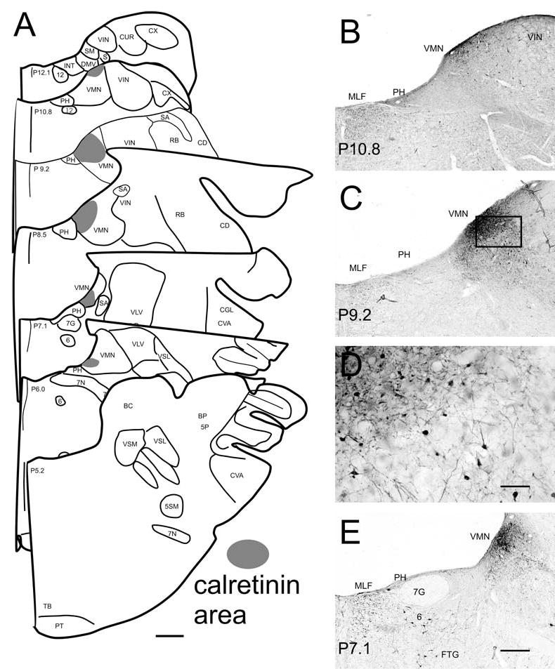
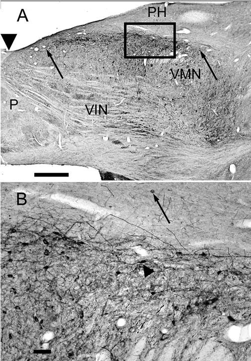
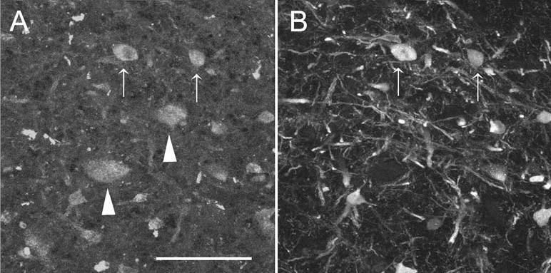
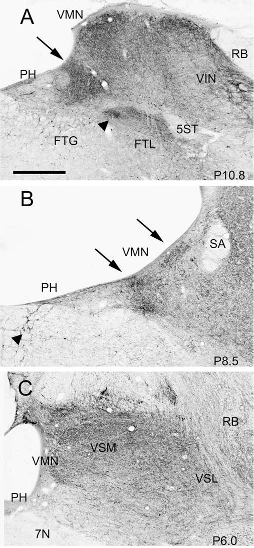
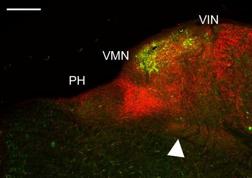
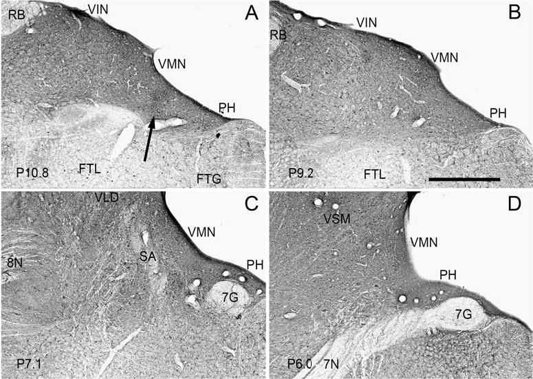
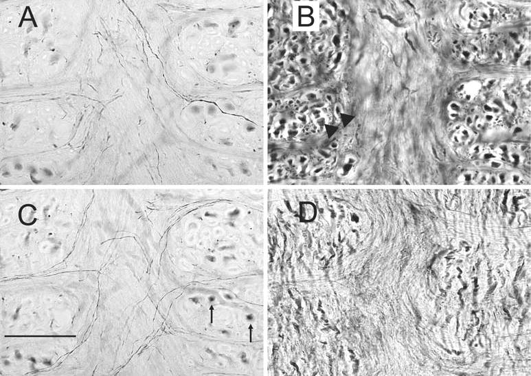
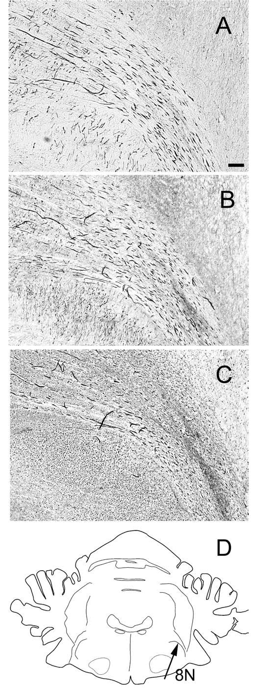
The vestibular nuclear complex (VNC) is classically divided into four nuclei on the basis of cytoarchitectonics. However, anatomical data on the distribution of afferents to the VNC and the distribution of cells of origin of different efferent pathways suggest a more complex internal organization. Immunoreactivity for calcium-binding proteins has proven useful in many areas of the brain for revealing structure not visible with cell, fiber or Golgi stains. We have looked at the VNC of the cat using immunoreactivity for the calcium-binding proteins calbindin, calretinin and parvalbumin. Immunoreactivity for calretinin revealed a small, intensely stained region of cell bodies and processes just beneath the fourth ventricle in the medial vestibular nucleus. A presumably homologous region has been described in rodents. The calretinin-immunoreactive cells in this region were also immunoreactive for choline acetyltransferase. Evidence from other studies suggests that the calretinin region contributes to pathways involved in eye movement modulation but not generation. There were focal dense regions of fibers immunoreactive to calbindin in the medial and inferior nuclei, with an especially dense region of label at the border of the medial nucleus and the nucleus prepositus hypoglossi. There is anatomical evidence that suggests that the likely source of these calbindin-immunoreactive fibers is the flocculus of the cerebellum. The distribution of calbindin-immunoreactive fibers in the lateral and superior nuclei was much more uniform. Immunoreactivity to parvalbumin was widespread in fibers distributed throughout the VNC. The results suggest that neurochemical techniques may help to reveal the internal complexity in VNC organization.


Figures

Fig. 1
Fig. 1
A The dense calretinin-immunoreactive area (shaded) shown on drawings of seven frontal sections through the vestibular nuclear complex from about P12 to P5 based on Berman (1968). B, C, E The calretinin area shown in photomicrographs on three frontal sections at different A-P levels. Also visible are scattered labeled cells outside this region in the VMN (B, C, D), labeled cells in VIN (B, C), labeled fibers in the MLF (B, C, E), and labeled cells in PH (B, C), and the abducens nucleus and reticular formation (E). The numbers at the bottom left of each panel show the approximate stereotaxic level of the section. D Higher magnification photomicrograph of the boxed region in C, showing the difference between the more dense central region, upper left, and more lightly labeled surrounding region, lower right. Both regions are included in the shaded areas in the schematics in A. Scale bars: A 1 mm, D 200 μm, B, C, E 500 μm
Fig. 2
Fig. 2
A The calretinin area in the VMN on a horizontal section at about −3.4. The anterior and posterior limits of the calretinin area are indicated by arrows. Note scattered labeled cells in the bordering nuclei VIN and PH. Arrowhead indicates an A-P level of about P14. B Higher magnification photomicrograph of the boxed area in A showing cells and processes in the calretinin area; the arrowhead points to a cell with a fusiform cell body. The arrow indicates a labeled cell in PH. Note the anterior-posterior orientation of the longer dendrites. P is posterior. Scale bars A 1 mm, B 100 μm
Fig. 3
Fig. 3
Colocalization of calretinin and ChAT in cells in the calretinin-immunoreactive area. A ChAT-immunoreactive cell bodies in the calretinin area at about P10. The arrows show two cell bodies that are also calretinin-immunoreactive; the arrowheads show cells which are not. B Calretinin-immunoreactive cells and fibers in the calretinin area. The arrows indicate the same cells shown in A. Scale bar: 50 μm
Fig. 4
Fig. 4
Changes in the pattern of calbindin-immunoreactivity in the VNC at different A-P levels, shown on three photomicrographs. The stereotaxic location of each section is indicated in the bottom right of each panel. A Arrow shows the trapezoidal calbindin-immunoreactive dense patch; arrowhead shows a small region of dense label in the reticular formation. Also visible are denser labeling along the ventricle in VMN and VIN. B The arrow on the left shows the two dense patches of label at the border of VMN and PH, and the arrow on the right the dense label along the ventricle. Arrowhead shows large calbindin-immunoreactive cell bodies embedded in the MLF. C Relatively uniform label in VMN and the VSM and VSL. Scale bar: 500 μm
Fig. 5
Fig. 5
Relative distributions of calretinin-immunoreactivity (green) and calbindin-immunoreactivity (red) on a section at about P11, at about the same level illustrated in Fig. 4A. Calretinin label is primarily in cells; calbindin label primarily in fibers. Arrowhead points to the dense calbindin cell label in the reticular formation. Scale bar: 500 μm
Fig. 6
Fig. 6
Widespread and uniform parvalbumin-immunoreactivity, predominantly in fibers and puncta, in all nuclei of the VNC shown on four photomicrographs at the stereotaxic levels indicated in the lower left corner of each panel. A Label in VMN, VIN and PH. Arrow shows the darker patch of label at the border of VMN and PH. B Uniform label in VMN, VIN and PH. C Uniform label in the VMN and VLD. D Uniform label in the VMN and VSM. Scale bar: 1 mm
Fig. 7
Fig. 7
Immunoreactivity for each of the calcium-binding proteins in and around the MLF; all sections are at about P9. A, C Calretinin-immunoreactivity shown in two photomicrographs of the same location at different planes of focus. There are fine-labeled fibers crossing the midline, and large stained fibers in the MLF cut in cross-section (arrows). B Parvalbumin-immunoreactive fibers in the MLF showing the densely-stained large diameter fibers (arrowheads) cut in cross-section. D Calbindin-immunoreactivity in the MLF; a few fine fibers cross the MLF, and there are short stained fibers with a dorsal-ventral orientation. Scale bar: 100 μm
Fig. 8
Fig. 8
Immunoreactivity for calcium-binding proteins in the fibers of the eighth nerve as it enters the brainstem, shown on photomicrographs of neighboring sections at about P7.0. A Calretinin-immunoreactive fibers. B Calbindin-immunoreactive fibers. C Parvalbumin-immunoreactive fibers. D The drawing shows an outline of a section through the brainstem at P7.

Similar articles

Cited by

References

    1. Adams JC. Heavy metal enhancement of DAB-based HRP reaction product. J Histochem Cytochem. 1981;29:775. - PubMed
    1. Akaike T. Neuronal organization of the vestibulospinal system in the cat. Brain Res. 1983;259:217–227. - PubMed
    1. Angaut P, Brodal A. The projection of the “vestibulocerebellum” onto the vestibular nuclei in the cat. Arch Ital Biol. 1967;105:441–479. - PubMed
    1. Arai R, Winsky L, Arai M, Jacobowitz DM. Immunohistochemical localization of calretinin in the rat hindbrain. J Comp Neurol. 1991;310:21–44. - PubMed
    1. Baimbridge KG, Celio MR, Rogers JH. Calcium-binding proteins in the nervous system. Trends Neurosci. 1992;15:303–308. - PubMed

Publication types